. 2019 Apr 16;9(21):11882-11893.
doi: 10.1039/c9ra00408d. eCollection 2019 Apr 12.

Biofunctionalization of decellularized porcine aortic valve with OPG-loaded PCL nanoparticles for anti-calcification

Affiliations

Biofunctionalization of decellularized porcine aortic valve with OPG-loaded PCL nanoparticles for anti-calcification

Yang Li et al. RSC Adv. .

Abstract

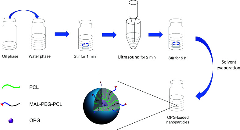
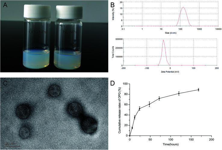
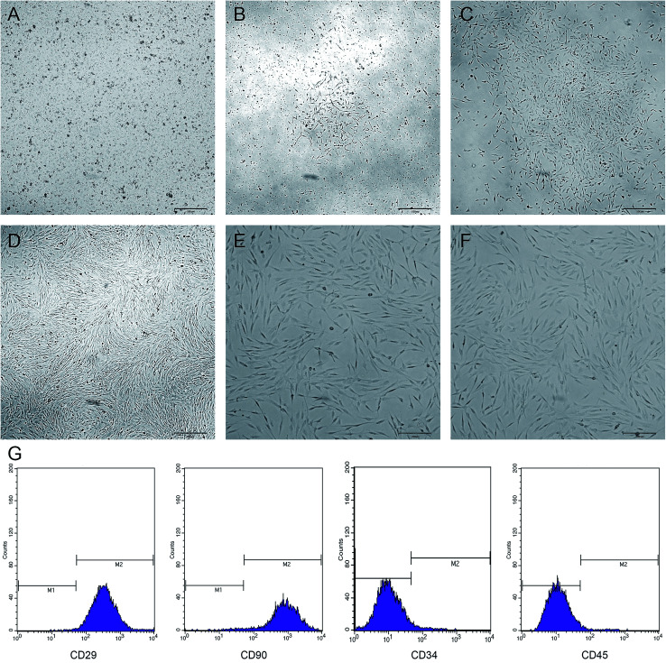
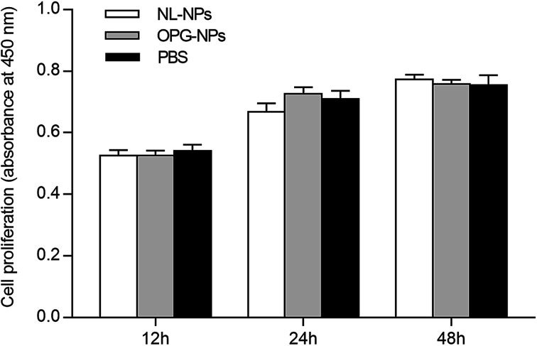
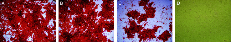
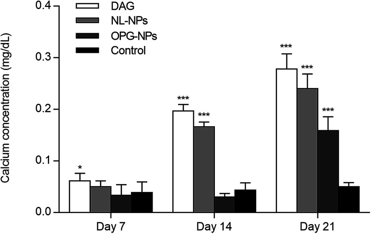
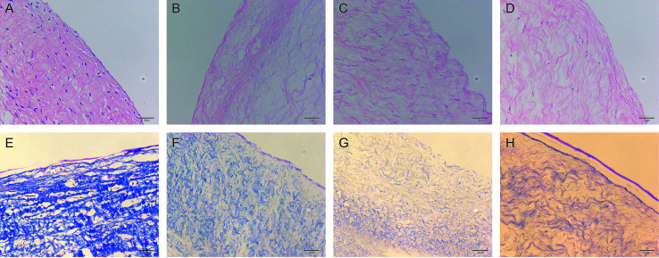
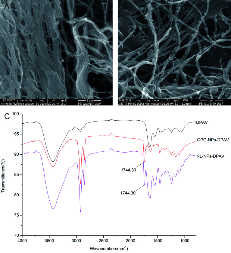
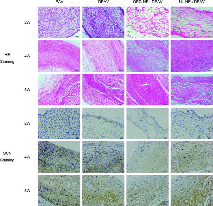
Decellularized valve stents are widely used in tissue-engineered heart valves because they maintain the morphological structure of natural valves, have good histocompatibility and low immunogenicity. However, the surface of the cell valve loses the original endothelial cell coverage, exposing collagen and causing calcification and decay of the valve in advance. In this study, poly ε-caprolactone (PCL) nanoparticles loaded with osteoprotegerin (OPG) were bridged to a decellularized valve using a nanoparticle drug delivery system and tissue engineering technology to construct a new anti-calcification composite valve with sustained release function. The PCL nanoparticles loaded with OPG were prepared via an emulsion solvent evaporation method, which had a particle size of 133 nm and zeta potential of -27.8 mV. Transmission electron microscopy demonstrated that the prepared nanoparticles were round in shape, regular in size, and uniformly distributed, with an encapsulation efficiency of 75%, slow release in vitro, no burst release, no cytotoxicity to BMSCs, and contained OPG nanoparticles in vitro. There was a delay in the differentiation of BMSCs into osteoblasts. The decellularized valve modified by nanoparticles remained intact and its collagen fibers were continuous. After 8 weeks of subcutaneous implantation in rats, the morphological structure of the valve was almost complete, and the composite valve showed anti-calcification ability to a certain extent.


Conflict of interest statement

There are no conflicts to declare.

Figures

Fig. 1
Fig. 1. Preparation of OPG-loaded nanoparticles via the emulsification solvent evaporation method.
Fig. 2
Fig. 2. Characterization of the nanoparticles: (A) OPG-loaded nanoparticles and non-loaded nanoparticles solution, (B) particle size and zeta potential distribution of OPG-loaded nanoparticles, (C) scanning electron microscopy image of OPG-loaded nanoparticles and (D) in vitro release of OPG-loaded nanoparticles.
Fig. 3
Fig. 3. Morphological features and cell surface markers of BMSCs: (A–D) cell morphology of primary culture for 1, 3, 5, and 7 days; (E and F) cell morphology of passages 2 and 3 and (G) cell surface markers of BMSCs, respectively.
Fig. 4
Fig. 4. Effects of OPG-loaded nanoparticles and non-loaded OPG nanoparticles on BMSC proliferation for 12 h, 24 h and 48 h.
Fig. 5
Fig. 5. Alizarin red staining (×40): (A) cells were treated with DAG and PBS, (B) cells were treated with DAG and NL-NPs, (C) cells were treated with DAG and OPG-NPs and (D) cells were treated with complete culture medium.
Fig. 6
Fig. 6. Quantitative determination of matrix calcification for 7, 14, and 21 days (*P < 0.05, ***P < 0.001).
Fig. 7
Fig. 7. HE staining and Masson's trichrome stain results of different groups (×400): (A and E) PAV group, (B and F) DPAV group, (C and G) OPG-NPs–DPAV group and (D and H) NL-NPs–DPAV group, respectively.
Fig. 8
Fig. 8. Characterization of OPG-loaded nanoparticles immobilized onto DPAV: (A and B) SEM image of DPAV and OPG-NPs–DPAV and (C) FTIR spectra of DPAV, NL-NPs–DPAV and OPG-NPs–DPAV groups.
Fig. 9
Fig. 9. HE staining and osteocalcin immunostaining results of the different groups after subdermal implantation for 2, 4 and 8 weeks. Images were captured under 200 magnified visual field.

Similar articles

Cited by

References

    1. Hisamatsu T. Miura K. Fujiyoshi A. Kadota A. Miyagawa N. Satoh A. Zaid M. Yamamoto T. Horie M. Ueshima H. Atherosclerosis. 2018;273:145–152. doi: 10.1016/j.atherosclerosis.2018.03.035. - DOI - PMC - PubMed
    1. Nkomo V. T. Gardin J. M. Skelton T. N. Gottdiener J. S. Scott C. G. Enriquez-Sarano M. Lancet. 2006;368:1005–1011. doi: 10.1016/S0140-6736(06)69208-8. - DOI - PubMed
    1. Brennan J. M. Edwards F. H. Zhao Y. O'Brien S. Booth M. E. Dokholyan R. S. Douglas P. S. Peterson E. D. Circulation. 2013;127:1647–1655. doi: 10.1161/CIRCULATIONAHA.113.002003. - DOI - PubMed
    1. Usprech J. Chen W. L. Simmons C. A. Wiley Interdiscip. Rev.: Syst. Biol. Med. 2016;8:169–182. - PubMed
    1. Zhu A. S. Grande-Allen K. J. Curr. Opin. Biomed. Eng. 2018;5:35–41. doi: 10.1016/j.cobme.2017.12.006. - DOI