. 2019 Apr 16;9(21):11810-11817.
doi: 10.1039/c9ra00617f. eCollection 2019 Apr 12.

Exposure to microwave irradiation at constant culture temperature slows the growth of Escherichia coli DE3 cells, leading to modified proteomic profiles

Affiliations

Exposure to microwave irradiation at constant culture temperature slows the growth of Escherichia coli DE3 cells, leading to modified proteomic profiles

Sina Atrin Mazinani et al. RSC Adv. .

Abstract

Despite a few decades of research, interest continues in understanding the potential influences of low energy microwave irradiation on biological systems. In the present study, growth of E. coli DE3 in LB media slowed in the presence of microwave irradiation (max. 10 W) while the temperature of cultures was maintained at 37 °C. Viable cell counts in microwave-irradiated cultures were also significantly lower. When microwave irradiation was ceased, E. coli growth was restored. A top-down proteomic analysis of total proteins isolated from control and microwave-irradiated E. coli cultures revealed differential abundance of 10 resolved protein spots, with multiple proteins identified in each following mass spectrometric analysis. Among these proteins, a number are involved in metabolism, suggesting alterations to metabolic activities following microwave irradiation. Furthermore, four amino acid-tRNA ligases were also identified, pointing to the possibility of stress responses in E. coli under microwave irradiation.


Conflict of interest statement

The authors declare no conflict of interest.

Figures

Fig. 1
Fig. 1. E. coli culture setup in a CEM microwave reactor.
Fig. 2
Fig. 2. Growth curves of E. coli. ■: cultured at 37 °C in a shaking incubator at 260 rpm; ●: cultured with stirring at 37 °C in a tube in the presence of microwave irradiation; ▲: cultured with stirring in a tube at 37 °C in an oil-bath in the absence of microwave exposure. All experiments were an average of biological triplicates. *P < 0.05; **P < 0.01 calculated for OD600 of cultures treated with microwave and control in oil bath.
Fig. 3
Fig. 3. Recovery of E. coli growth after ceasing microwave exposure at 5 h as indicated by the decreased difference in OD600. Both control and microwave-treated cultures were carried out in biological triplicates.
Fig. 4
Fig. 4. Viable cell counts 21 h after microwave exposure as compared with control. Graph shows mean ± std. dev.
Fig. 5
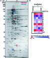
Fig. 5. Representative 2DE gel used for the analysis of proteins extracted from control and microwave-treated E. coli cultures. (A) Annotated 2DE gel image showing protein spots which changed in abundance following microwave irradiation. Spots which decreased in abundance are delineated in red; those which increased are delineated in blue. Refer to Table 1 for calibrated pI and molecular weights of each delineated spot. (B) Heat map displaying normalised spot volume ratios in irradiated cultures relative to control cultures, across three biological replicates, averaged from three technical gel replicates each. Mean ratio is also shown.
Fig. 6
Fig. 6. Setup for DO measurement. LB media isolated from the environment with the use of a foam plug were cooled through circulation in an internal cooling tube.

Similar articles

Cited by

References

    1. Kuhnert N. Angew. Chem., Int. Ed. 2002;41:1863. doi: 10.1002/1521-3773(20020603)41:11<1863::AID-ANIE1863>3.0.CO;2-L. - DOI - PubMed
    1. de la Hoz A. Diaz-Ortiz A. Moreno A. Chem. Soc. Rev. 2005;34:164. doi: 10.1039/B411438H. - DOI - PubMed
    1. Dudley G. B. Stiegman A. E. Rosana M. R. Angew. Chem., Int. Ed. 2013;52:7918. doi: 10.1002/anie.201301539. - DOI - PubMed
    1. Gutmann B. Schwan A. M. Reichart B. Gspan C. Hofer F. Kappe C. O. Angew. Chem., Int. Ed. 2011;50:7636. doi: 10.1002/anie.201100856. - DOI - PubMed
    1. Kappe C. O. Angew. Chem., Int. Ed. 2013;52:7924. doi: 10.1002/anie.201304368. - DOI - PubMed