. 2020 Sep 30;10(59):35765-35775.
doi: 10.1039/d0ra06223e. eCollection 2020 Sep 28.

A sandwich-type bacteriophage-based amperometric biosensor for the detection of Shiga toxin-producing Escherichia coli serogroups in complex matrices

Affiliations

A sandwich-type bacteriophage-based amperometric biosensor for the detection of Shiga toxin-producing Escherichia coli serogroups in complex matrices

Irwin A Quintela et al. RSC Adv. .

Abstract

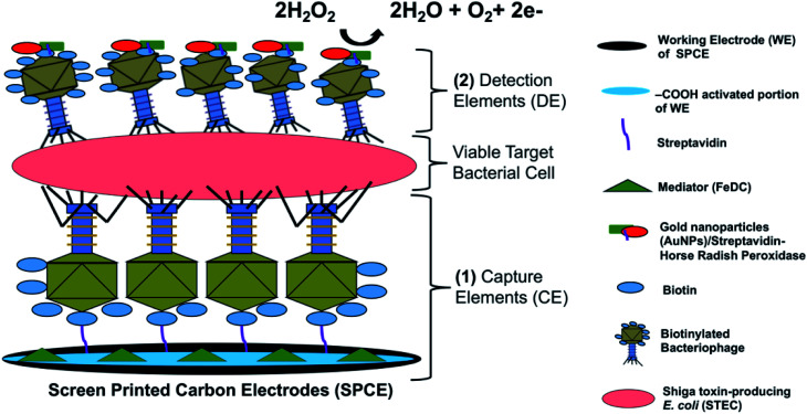
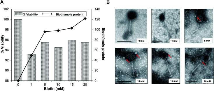
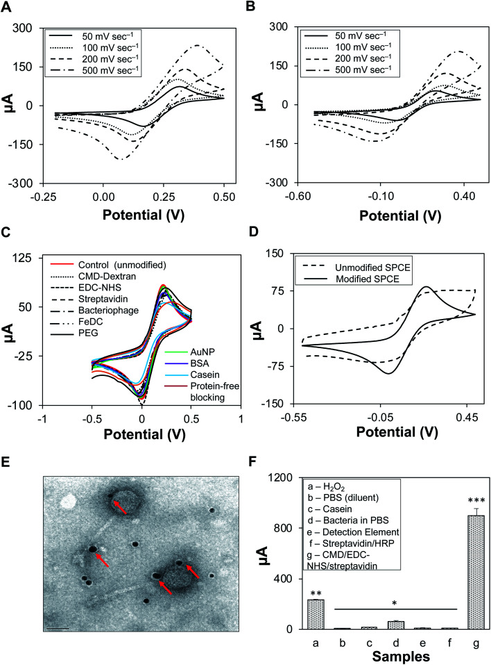
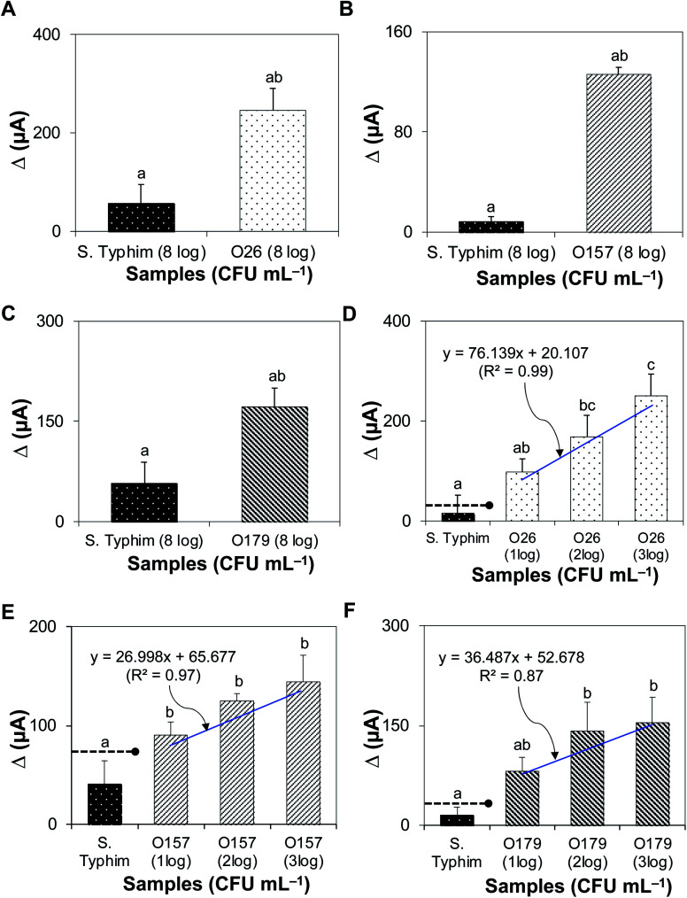
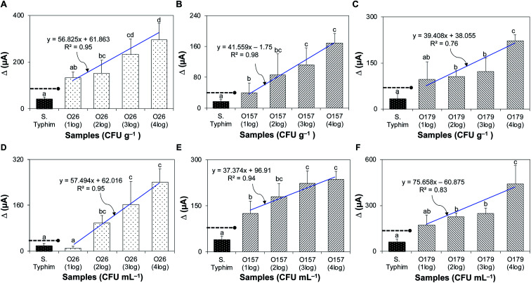
Immuno-based biosensors are a popular tool designed for pathogen screening and detection. The current antibody-based biosensors employ direct, indirect, or sandwich detection approaches; however, instability, cross-reactivity, and high-cost render them unreliable and impractical. To circumvent these drawbacks, here we report a portable sandwich-type bacteriophage-based amperometric biosensor, which is highly-specific to various Shiga toxin-producing Escherichia coli (STEC) serogroups. Environmentally isolated and biotinylated bacteriophages were directly immobilized onto a streptavidin-coated screen-printed carbon electrode (SPCE), which recognized and captured viable target cells. Samples (50 μL) were transferred to these bacteriophage-functionalized SPCEs (12 min, room temp) before sequentially adding a bacteriophage-gold nanoparticle solution (20 μL), H2O2 (40 mM), and 1,1'-ferrocenedicarboxylic acid for amperometric tests (100 mV s-1) and analysis (ANOVA and LSD, P < 0.05). The optimum biotin concentration (10 mM) retained 94.47% bacteriophage viability. Non-target bacteria (Listeria monocytogenes and Salmonella Typhimurium) had delta currents below the threshold of a positive detection. With less than 1 h turn-around time, the amperometric biosensor had a detection limit of 10-102 CFU mL-1 for STEC O157, O26, and O179 strains and R 2 values of 0.97, 0.99, and 0.87, respectively, and a similar detection limit was observed in complex matrices, 10-102 CFU g-1 or mL-1 with R 2 values of 0.98, 0.95, and 0.76, respectively. The newly developed portable amperometric biosensor was able to rapidly detect viable target cells at low inoculum levels, thus providing an inexpensive and improved alternative to the current immuno- and laboratory-based STEC screening methods.


Conflict of interest statement

There are no conflicts to declare.

Figures

Fig. 1
Fig. 1. The detection principle of the bacteriophage-based functionalized SPCE biosensor. Two sets of highly specific biotinylated bacteriophages were used as (1) SPCE-bound capture element and (2) AuNP–S-HRP-tagged detection element binding to captured viable STEC cells for peroxidase-coupled SPCE redox reactions.
Fig. 2
Fig. 2. Biotinylation and viability of STEC O179 bacteriophage. (A) The effects of biotinylation in relation to bacteriophage viability. (B) TEM images of biotinylated bacteriophages with increasing biotin concentrations (0 mM–20 mM) and coupling it with streptavidin-coated QDots. Scale: 100 nm.
Fig. 3
Fig. 3. Development and optimization of capture and detection elements. K3[Fe(CN6)] (0.5 mM) was used with (A) 0.1 M sulfuric acid or (B) 1× PBS as a supporting electrolyte at increasing scan rates. (C) Cyclic voltammograms (100 mV s−1) of chemical reagents. (D) Cyclic voltammogram of unmodified and modified SPCEs. (E) TEM image of the detection element solution, AuNPs (signal amplifiers) seen as dark spots (indicated by arrows) were bound to biotinylated STEC O179 bacteriophages. (F) Determination of sources of background noise by amperometry. Background signal approximately 900 μA (896 ± 58.24 μA) was determined and used as the baseline value to analyze subsequent amperometric tests. Bars with different asterisks (*) are significantly different (P < 0.05).
Fig. 4
Fig. 4. Specificity and sensitivity of the biosensor in a pure culture set up. (A–C) The specificity of the assay was challenged by testing and comparing Δ currents between target STEC bacteria and non-target samples (S. Typhimurium). (D–F) The sensitivity of the assay was tested from 10 to 103 CFU mL−1 of target STEC (O26, O157, and O179). The threshold of (+) detection was defined by the signal-to-noise (S/N) characteristics as S/N > 2. Dashed lines with solid circles indicate the threshold for positive detection. Bars with different lower-case letters are significantly different (P < 0.05). The detection limit was determined by the statistical significance of Δ current between non-target bacteria and the lowest inoculum of target bacteria that had Δ current above the signal threshold for positive detection.
Fig. 5
Fig. 5. Detection of STEC strains in artificially inoculated complex matrices. (A–C) Bar graphs showing the sensitivity of detection for STEC O26, O157, and O179 in the fresh ground beef matrix. (D–F) Bar graphs showing the sensitivity of detection for STEC O26, O157, and O179 in pasteurized apple juice. Dashed lines with solid circles indicate the threshold for positive detection. Bars with different lower-case letters are significantly different (P < 0.05). The detection limit was determined by the statistical significance of Δ current between non-target bacteria and the lowest inoculum of target bacteria that had Δ current above the signal threshold for positive detection.
Fig. 6
Fig. 6. Detection of STEC strains in natural environmental water samples. The natural environmental water samples were collected at four different sites. Bar graphs showing Δ currents (μA) for (A) STEC O26, (B) STEC O157, and (C) STEC O179. Dashed lines with solid circle indicate the threshold for positive detection established in pure culture setup.

Similar articles

Cited by

References

    1. Vallières E. Saint-Jean M. Rallu F. J. Clin. Microbiol. 2013;51:481–486. doi: 10.1128/JCM.02219-12. - DOI - PMC - PubMed
    1. Beutin L. Fach P. Microbiol. Spectrum. 2014;2:3. - PubMed
    1. Quintela I. A. de los Reyes B. G. Lin C.-S. Wu V. C. Nanoscale. 2015;7:2417–2426. doi: 10.1039/C4NR05869K. - DOI - PubMed
    1. Fuller C. A. Pellino C. A. Flagler M. J. Strasser J. E. Weiss A. A. Infect. Immun. 2011;79:1329–1337. doi: 10.1128/IAI.01182-10. - DOI - PMC - PubMed
    1. Mayer C. L. Leibowitz C. S. Kurosawa S. Stearns-Kurosawa D. J. Toxins. 2012;4:1261–1287. doi: 10.3390/toxins4111261. - DOI - PMC - PubMed

LinkOut - more resources