. 2020 Sep 30;10(59):35753-35764.
doi: 10.1039/d0ra05901c. eCollection 2020 Sep 28.

Iron oxide nanoparticle-induced hematopoietic and immunological response in rats

Affiliations

Iron oxide nanoparticle-induced hematopoietic and immunological response in rats

Usha Singh Gaharwar et al. RSC Adv. .

Abstract

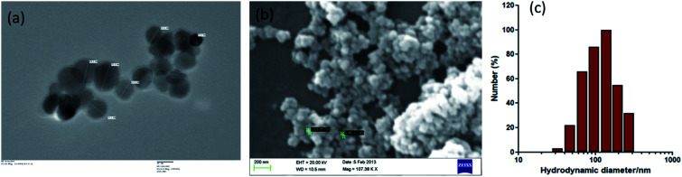
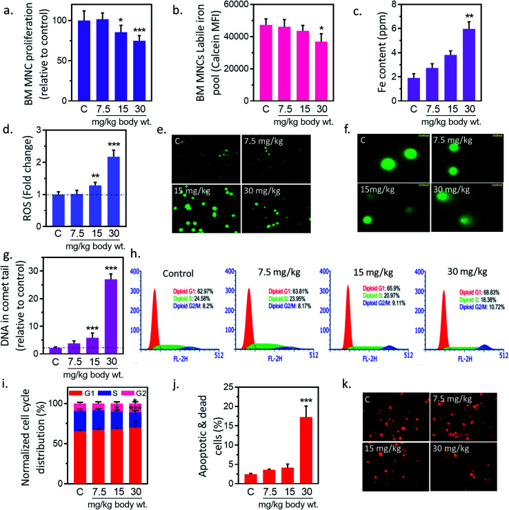
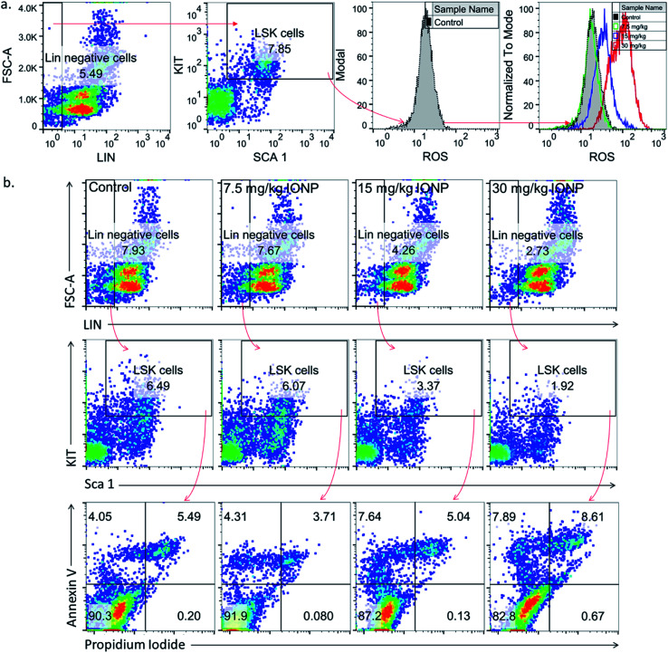
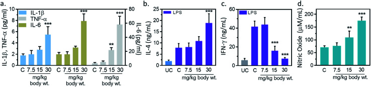
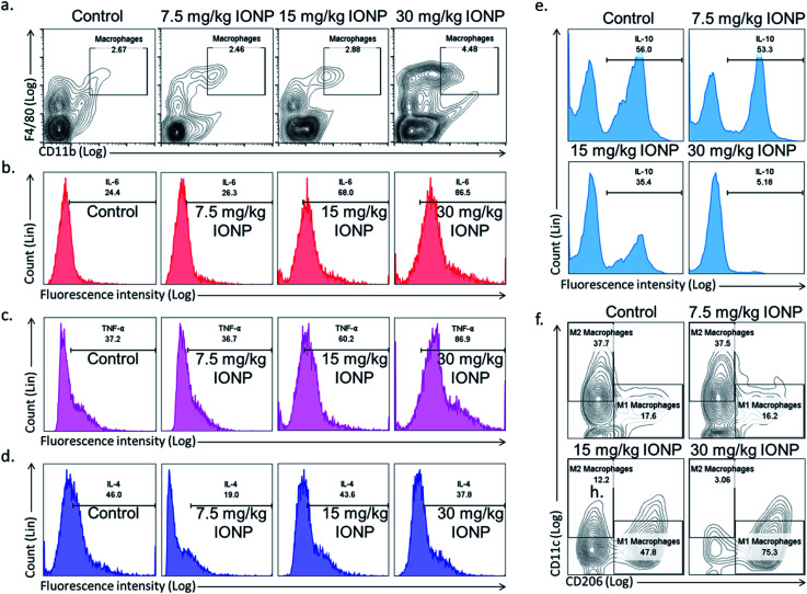
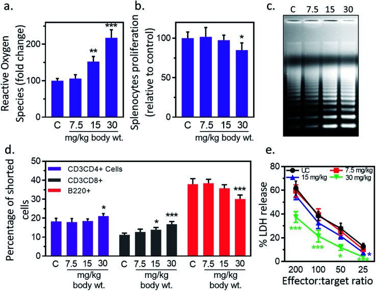
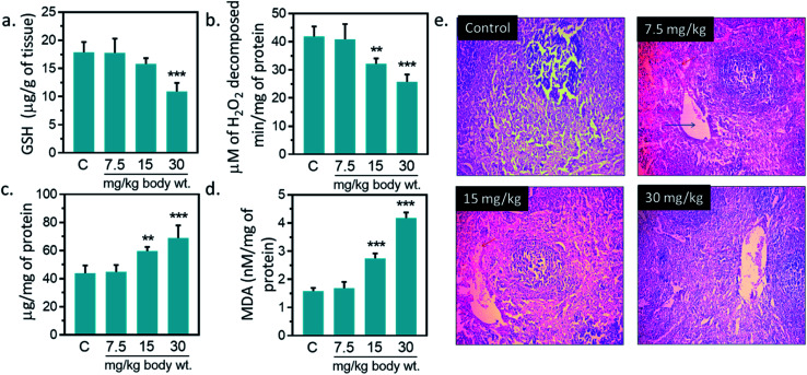
The application and use of iron oxide nanoparticless (IONPs) in the biomedical field are steadily increasing, although it remains uncertain whether IONPs are safe or should be used with caution. In the present study, we investigated the toxicity profile of ultrafine IONPs in rats administered with 7.5, 15 and 30 mg IONPs/kg body wt intravenously once a week for 4 weeks. IONP treatment reduces bone marrow-mononuclear cell proliferation, increases free radical species and DNA damage leading to growth arrest and subsequently apoptosis induction at 15 and 30 mg doses. It also induces apoptosis in undifferentiated hematopoietic stem cells. IONP treatment significantly increased the pro-inflammatory cytokine (Interleukin (IL)-1β, TNF-α, and IL-6) level in serum. The induction in inflammation was likely mediated by splenic M1 macrophages (IL-6 and TNF-α secretion). IONP treatment induces splenocyte apoptosis and alteration in the immune system represented by reduced CD4+/CD8+ ratio and increased B cells. It also reduces innate defense represented by lower natural killer cell cytotoxicity. IONP administration markedly increased lipid peroxidation in the spleen, while the glutathione level was reduced. Similarly, superoxide dismutase activity was increased and catalase activity was reduced in the spleen of IONP-treated rats. At an organ level, IONP treatment did not cause any significant injury or structural alteration in the spleen. Collectively, our results suggest that a high dose of ultrafine IONPs may cause oxidative stress, cell death, and inflammation in a biological system.


Conflict of interest statement

There are no conflicts to declare.

Figures

Fig. 1
Fig. 1. Characterization of Iron oxide nanoparticles (IONPs). (a) Transmission electron microscope image of IONPs, scale bar: 20 nm; (b) scanning electron microscope image of IONPs. (c) Dynamic light scattering (DLS) histogram for hydrodynamic size distribution of IONPs.
Fig. 2
Fig. 2. Iron nanoparticles (IONPs) induce cytotoxic and apoptotic response in bone marrow mononuclear cells (BM-MNCs) of rats. (a) BM-MNCs proliferation, detected by MTT; (b) labile iron pool in BM-MNCs, detected by calcein; (c) iron level in BM-MNCs, detected by coupled plasma atomic emission spectroscopy; (d and e) relative change in BM-MNCs ROS level, detected by DC-FDA; (f and g) BM-MNCs DNA damage, detected by alkaline comet assay; (h and i) activation of cell cycle check point in BM-MNCs (bar represents normalized % distribution of cells in different cell cycle stage); (j) BM-MSCs apoptosis, detected by annexin V propidium iodide; (k) BM-MNCs mitochondrial membrane potential (ΔΨm), measured by Mitotracker CMXROS. Rats were treated with IONPs intravenously for 28 days in a 7 day interval. Each data point represents mean ± SD. n: 6. *p < 0.05; **p < 0.01; and ***p < 0.001 versus saline control.
Fig. 3
Fig. 3. Iron nanoparticles (IONPs) increase reactive oxygen species in immature bone marrow hematopoietic cells (lineage negative, Sca1 positive, c-kit negative, LSK cells). (a) Representative gating strategy for LSK BM populations and ROS level in different groups, measured by DCFDA using flow cytometry; (b) gating strategy for bone marrow LSK cells, LSK cell apoptosis in different treatment groups. Rats were treated with IONPs intravenously for 28 days in a 7 day interval.
Fig. 4
Fig. 4. Iron oxide nanoparticles (IONPs) induce inflammatory response. (a) Blood IL-1β, IL-6 and TNF-α after IONPs treatment; (b) IL-4 production in different treatment groups; (c) IFN-γ production in different treatment groups; (d) nitric oxide production in different treatment groups. All graphs present average ± SEM. n: 6. Rats were treated with IONPs intravenously for 28 days in a 7 days interval. **p < 0.01; and ***p < 0.001 versus saline control.
Fig. 5
Fig. 5. IONPs induces inflammatory response by macrophage polarization. (a) Characterization of rat splenic macrophages (F4/80highCD11bhigh) by flow cytometry; (b) IL-6 secretion by macrophages (F4/80highCD11bhigh); (c) TNF-α secretion by macrophages (F4/80highCD11bhigh); (d) IL-4 secretion by macrophages (F4/80highCD11bhigh); (e) IL-10 secretion by macrophages (F4/80highCD11bhigh); (f) splenic M1 (F4/80highCD11bhighCD11c) and M2 macrophages (F4/80highCD11bhighCD206) proportion by flow cytometry. Rats were treated with IONPs intravenously for 28 days in a 7 day interval.
Fig. 6
Fig. 6. Iron oxide nanoparticles (IONPs) induce splenocytes apoptotic response. (a) IONPs treatment increases free radicals in splenocytes, measured by DC-FDA assay; (b) IONPs treatment reduces splenocytes proliferation, measured by MTT assay; (c) IONPs treatment increased splenocytes apoptosis, measured by ladder assay; (d) IONPs exposure increased Tc and Th cells and reduces B cells in spleen; (e) NK cells cytotoxicity against YAC-1 cells. Rats were treated with IONPs intravenously for 28 days in a 7 day interval. Each data point represents mean ± SD. n: 6. *p < 0.05; **p < 0.01; and ***p < 0.001 versus saline control.
Fig. 7
Fig. 7. Effect of Iron oxide nanoparticles (IONPs) on anti-oxidative enzyme level in spleen and spleen architecture. (a) GSH content; (b) catalase activity; (c) superoxide dismutase activity; (d) MDA level; (e) Haematoxylin–eosin staining of spleen sections for seeing the IONPs intoxication. Each data point represents mean ± SD. n: 6. Photomicrograph represents the one of 6-independent experiment. Rats were treated with IONPs intravenously for 28 days in a 7 day interval. *p < 0.05; **p < 0.01; and ***p < 0.001 versus saline control.

Similar articles

Cited by

References

    1. Gaharwar U. S. Meena R. Rajamani P. Iron oxide nanoparticles induced cytotoxicity, oxidative stress and DNA damage in lymphocytes. J. Appl. Toxicol. 2017;37(10):1232–1244. doi: 10.1002/jat.3485. - DOI - PubMed
    1. Feng Q. Liu Y. Huang J. Chen K. Huang J. Xiao K. Uptake, distribution, clearance, and toxicity of iron oxide nanoparticles with different sizes and coatings. Sci. Rep. 2018;8(1):1–3. doi: 10.1038/s41598-017-17765-5. - DOI - PMC - PubMed
    1. Alphandéry E. Biodistribution and targeting properties of iron oxide nanoparticles for treatments of cancer and iron anemia disease. Nanotoxicology. 2019;13(5):573–596. doi: 10.1080/17435390.2019.1572809. - DOI - PubMed
    1. Gaharwar U. S. Meena R. Rajamani P. Biodistribution, Clearance and Morphological Alterations of Intravenously Administered Iron Oxide Nanoparticles in Male Wistar Rats. Int. J. Nanomed. 2019;14:9677. doi: 10.2147/IJN.S223142. - DOI - PMC - PubMed
    1. Singh N. Jenkins G. J. Asadi R. Doak S. H. Potential toxicity of superparamagnetic iron oxide nanoparticles (SPION) Nano Rev. 2010;1(1):5358. doi: 10.3402/nano.v1i0.5358. - DOI - PMC - PubMed