. 2020 May 12;10(31):18054-18061.
doi: 10.1039/d0ra01807d. eCollection 2020 May 10.

An ultra-sensitive T 2-weighted MR contrast agent based on Gd3+ ion chelated Fe3O4 nanoparticles

Affiliations

An ultra-sensitive T 2-weighted MR contrast agent based on Gd3+ ion chelated Fe3O4 nanoparticles

Jing Chen et al. RSC Adv. .

Abstract

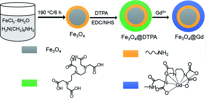
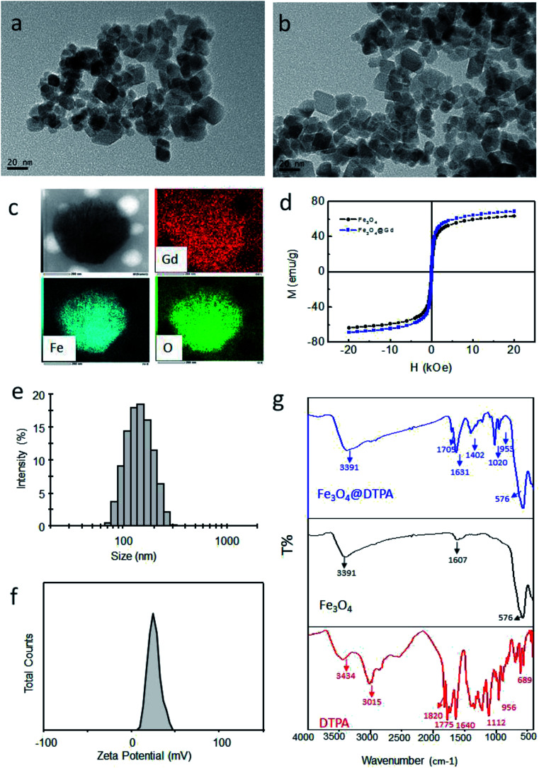
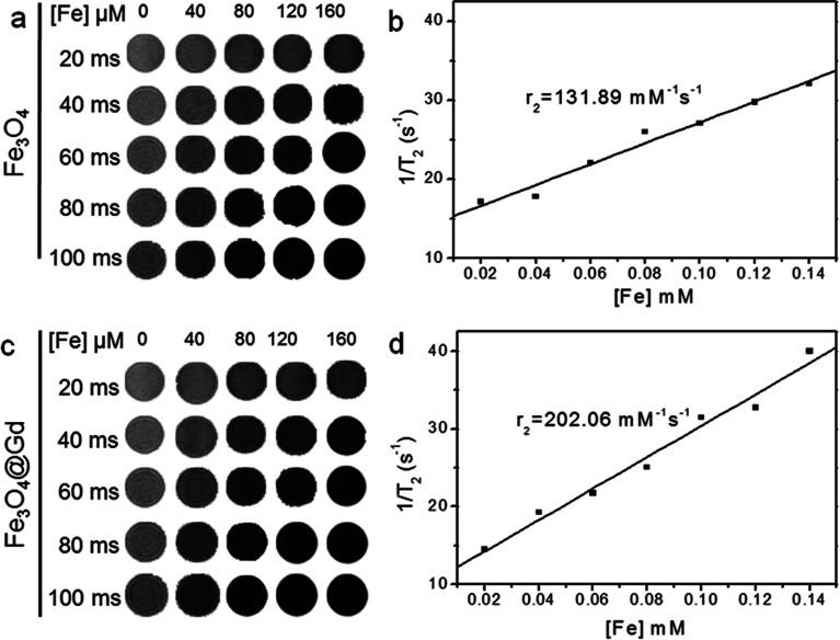
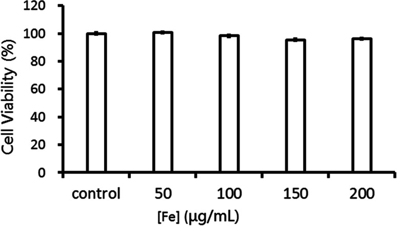
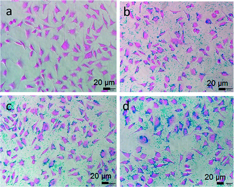
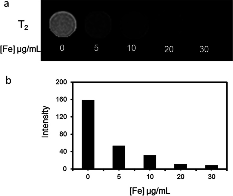
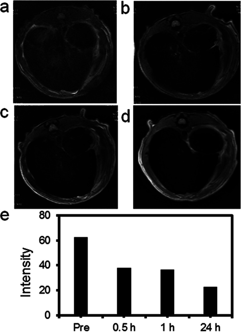
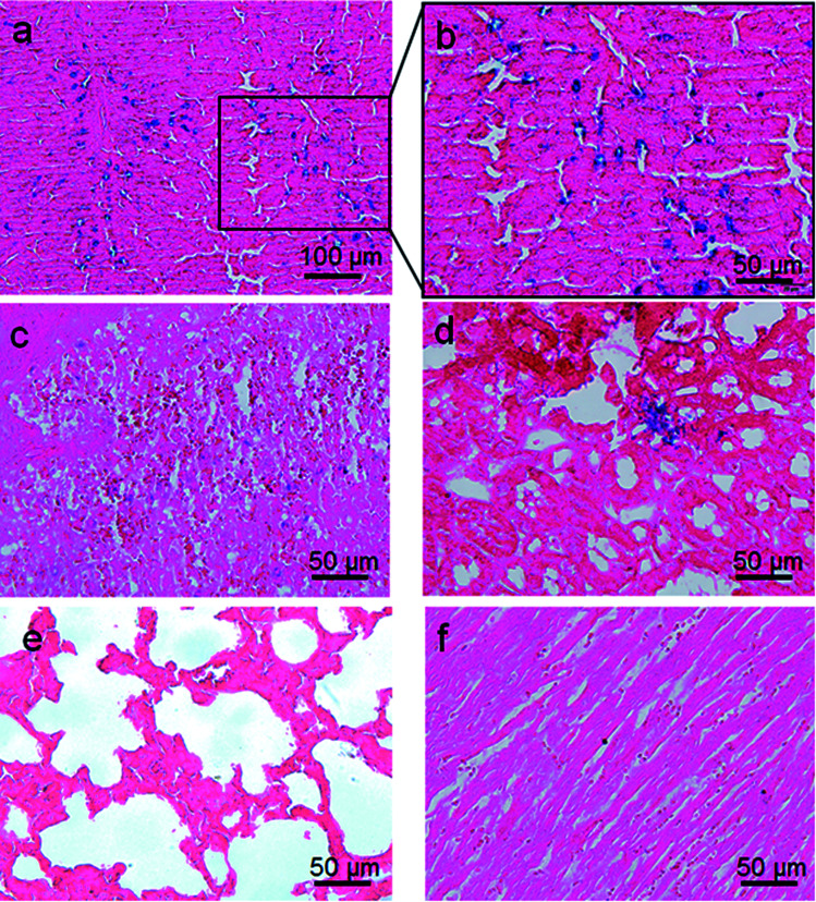
An ultra-sensitive T 2-weighted MR imaging contrast agent was prepared based on Fe3O4 nanoparticles and Gd3+ ions (Fe3O4@Gd). Amino modified Fe3O4 nanoparticles were conjugated to diethylenetriamine pentaacetic acid, and finally coordinated with Gd3+ ions. The nanoparticles had a uniform morphology with a size of 100 nm and a Gd/Fe mass ratio of 1/110. The r 2 (transverse relaxivity) of the Fe3O4 nanoparticles increased from 131.89 mM-1 s-1 to 202.06 mM-1 s-1 after coordination with Gd3+ ions. MR measurements showed that the aqueous dispersion of Fe3O4@Gd nanoparticles had an obvious concentration-dependent negative contrast enhancement. Hepatoma cells were selected to test the cytotoxicity and MR imaging effect. The application of Fe3O4@Gd nanoparticles as contrast agents was also exploited in vivo for T 2-weighted MR imaging of rat livers. All the results showed the effectiveness of the nanoparticles in MR diagnosis.


Conflict of interest statement

The authors declare no conflict of interest.

Figures

Scheme 1
Scheme 1. Schematic illustration of the formation of Fe3O4@Gd nanoparticles.
Fig. 1
Fig. 1. Characterization of nanoparticles. (a) TEM image of Fe3O4 nanoparticles; (b) TEM image of Fe3O4@Gd nanoparticles; (c) EDX mapping images of one nanoparticle; (d) the magnetic hysteresis loops of Fe3O4 and Fe3O4@Gd nanoparticles at room temperature; (e) size distribution of Fe3O4@Gd nanoparticles; (f) zeta potential of Fe3O4@Gd nanoparticles; (g) IR spectra of Fe3O4@DTPA nanoparticles, Fe3O4 nanoparticles and DTPA.
Fig. 2
Fig. 2. T 2-weighted MR images at different Fe concentration in 0.5% agarose gel at different TE time and the analysis of relaxation rate vs. Fe concentration. (a and b) Fe3O4 nanoparticles. (c and d) Fe3O4@Gd nanoparticles.
Fig. 3
Fig. 3. Cell viability of HepG2 cells incubated with Fe3O4@Gd nanoparticles at various Fe concentrations.
Fig. 4
Fig. 4. Prussian blue staining images of HepG2 cells incubated with Fe3O4@Gd nanoparticles at various iron concentration ((a) – control, (b) – 10 μg mL−1, (c) – 20 μg mL−1, (d) – 30 μg mL−1).
Fig. 5
Fig. 5. MR investigation in vitro. (a) T2-weighted MR images of HepG2 cells incubated with Fe3O4@Gd nanoparticles with iron concentration ranging from 0–30 μg mL−1 and (b) the corresponding analysis of MR signal intensity.
Fig. 6
Fig. 6. MR images of rat liver before and after vein tail injection of Fe3O4@Gd nanoparticles (10 mg kg−1). (a) Pre-injection; (b) 30 min after injection; (c) 60 min after-injection; (d) 24 h after injection; (e) the corresponding T2-weighted MR signal intensity of the liver.
Fig. 7
Fig. 7. Prussian blue staining images of the major organs of SD rats ((a and b) – liver, (c) – spleen, (d) – kidney, (e) – lung and (f) – heart). The sections were acquired from rat at 24 h after intravenous injection of 10 mg kg−1 Fe3O4@Gd nanoparticles.

Similar articles

Cited by

References

    1. Siegel R. L. Miller K. D. Jemal A. Ca-Cancer J. Clin. 2020;70:7–30. doi: 10.3322/caac.21590. - DOI - PubMed
    1. Gobbo O. Sjaastad K. Radomski M. Volkov Y. Prina-Mello A. Theranostics. 2015;5:1249–1263. doi: 10.7150/thno.11544. - DOI - PMC - PubMed
    1. Busquets M. Estelrich J. Sánchez-Martín M.-J. Int. J. Nanomed. 2015;10:1727. doi: 10.2147/IJN.S76501. - DOI - PMC - PubMed
    1. Weissleder R. Pittet M. Nature. 2008;452:580–589. doi: 10.1038/nature06917. - DOI - PMC - PubMed
    1. Boros E. Gale E. M. Caravan P. Dalton Trans. 2015;44:4804–4818. doi: 10.1039/C4DT02958E. - DOI - PMC - PubMed