. 2020 Jun 23;10(40):23908-23915.
doi: 10.1039/d0ra03803b. eCollection 2020 Jun 19.

Arrayed nanopore silver thin films for surface-enhanced Raman scattering

Affiliations

Arrayed nanopore silver thin films for surface-enhanced Raman scattering

Weiwei Zhang et al. RSC Adv. .

Abstract

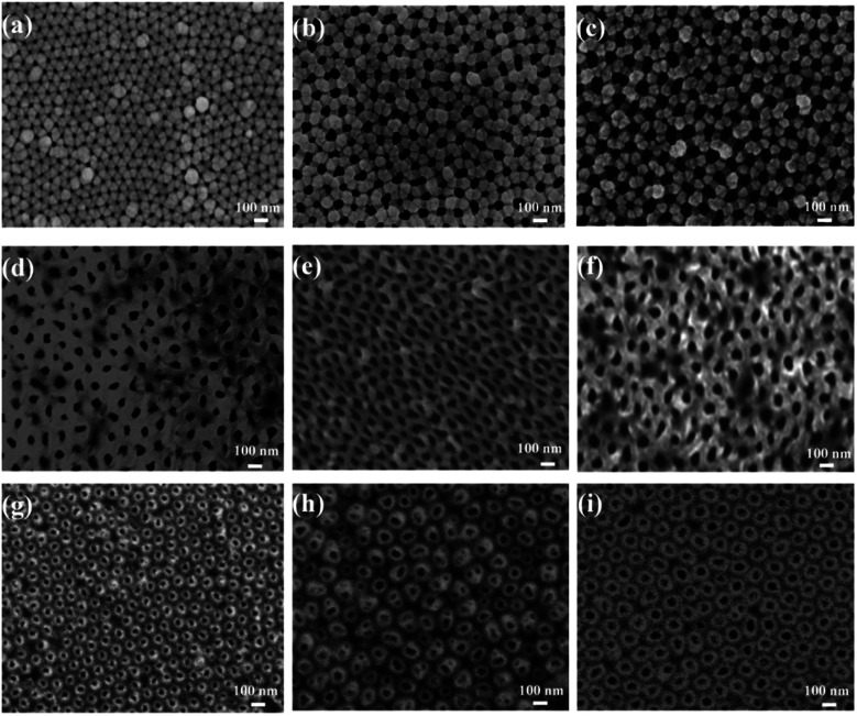
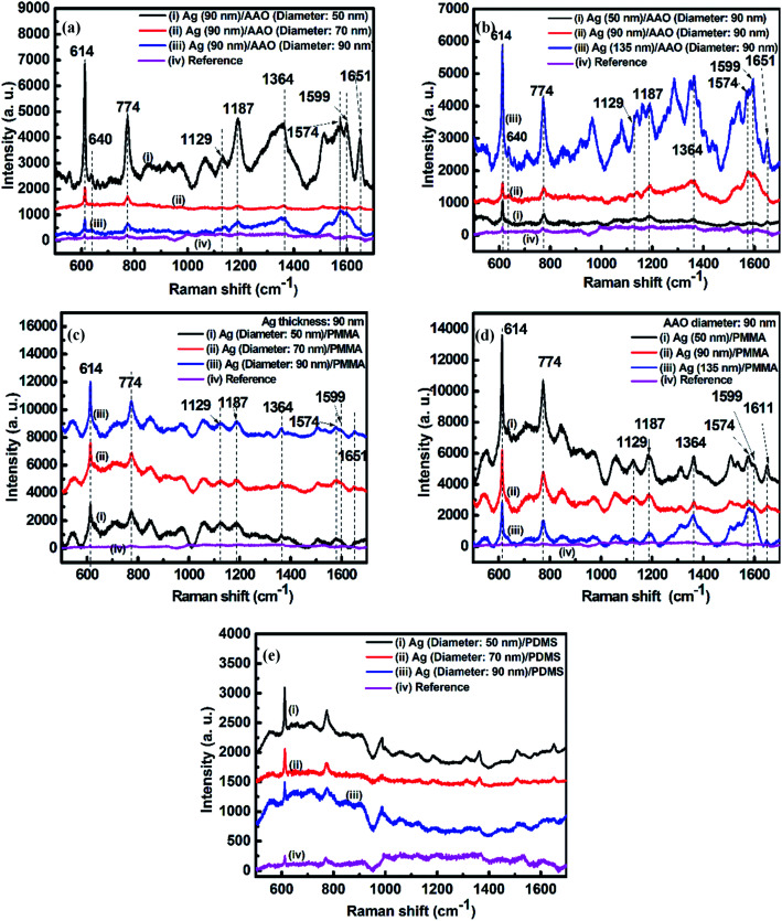
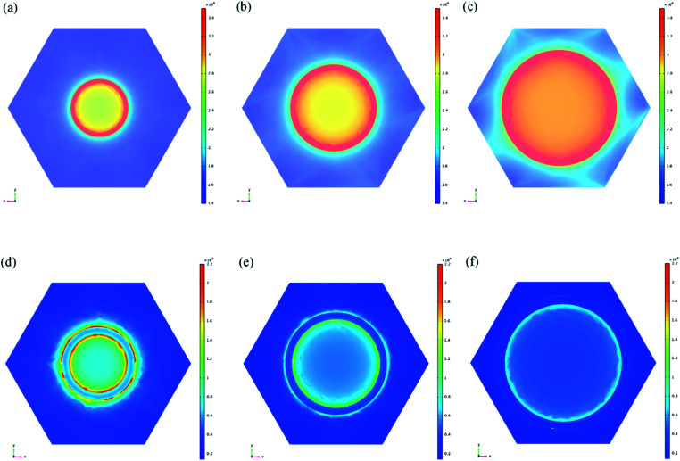
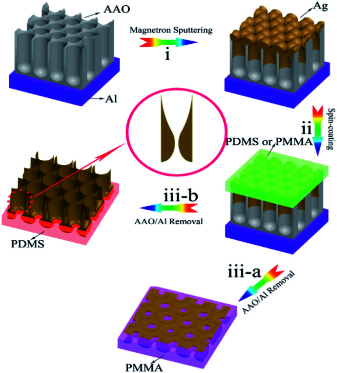
Active substrates are crucial for surface-enhanced Raman scattering (SERS). Among these substrates, large uniform area arrayed nanoporous silver thin films have been developed as active substrates. Arrayed nanoporous silver thin films with unique anisotropic morphologies and nanoporous structures can be fabricated onto the nanoporous anodic aluminum oxide (AAO) of controlled pore size and interspacing by precisely tuning the sputtering parameters. These thin films preserve locally enhanced electromagnetic fields by exciting the surface plasmon resonance, which is beneficial for SERS. In this study, nanoporous silver thin films were transferred into polymethylmethacrylate (PMMA) and polydimethylsiloxane (PDMS) substrates using our recently invented template-assisted sol-gel phase inverse-imprinting process to form two different nanopore thin films. The as-formed Ag nanoporous thin films on PMMA and PDMS exhibited intensively enhanced SERS signals using Rhodamine 6G (R6G) as the model molecule. The two nanopore thin films exhibited opposite pore size-dependent SERS tendencies, which were elucidated by the different enhancement tendencies of the electric field around pores of different diameters. In particular, the Ag nanoporous thin film on PMMA exhibited an R6G detection limit of as low as 10-6 mol L-1, and the SERS enhancement factor (EF) was more than 106. The low detection limit and large EF demonstrated the high sensitivity of the as-prepared SERS substrates for label-free detection of biomolecules. Compared with conventional smooth films, this nanopore structure can facilitate future application in biomolecular sensors, which allows the detection of single molecules via an electronic readout without requirement for amplification or labels.


Conflict of interest statement

There are no conflicts to declare.

Figures

Fig. 1
Fig. 1. SEM images of the nanoporous silver films with different pore sizes based on the AAO templates. The thickness of the silver film was 90 nm, and the pristine AAO pore diameters were (a) 50, (b) 70, and (c) 90 nm. SEM characterization of the nanoporous silver films based on PMMA substrates was assisted by the different pore diameters. The thickness of the silver film was 90 nm, and the pristine AAO pore diameters were (d) 50, (e) 70, and (f) 90 nm. SEM characterization of the nanoporous silver films based on PDMS substrates was assisted by the different pore diameters. The thickness of the silver film was 90 nm, and the pristine AAO pore diameters were (g) 50, (h) 70, and (i) 90 nm.
Fig. 2
Fig. 2. (a) SERS of R6G absorbed on 90 nm-thick Ag nanoporous films deposited on the AAO templates with diameters of (i) 50, (ii) 70, and (iii) 90 nm; (b) SERS of R6G absorbed on (i) 50, (ii) 90, and (iii) 135 nm-thick Ag nanoporous films deposited on the same AAO template with a diameter of 90 nm; (c) SERS of R6G absorbed on 90 nm-thick Ag films transferred from AAO templates that had diameters of (i) 50, (ii) 70, and (iii) 90 nm to PMMA substrates; (d) SERS of R6G absorbed on (i) 50, (ii) 90, and (iii) 135 nm-thick Ag nanoporous films transferred from the same AAO template with a diameter of 90 nm to the PMMA substrates; (e) SERS of R6G absorbed on 90 nm-thick Ag films transferred from AAO templates with diameters of (i) 50, (ii) 70, and (iii) 90 nm to PMMA substrates. Curves (iv) in (a)–(d) show the SERS signal of R6G/PMMA with a concentration of 10−2 mol L−1. Curve (iv) in (e) shows the SERS signal of R6G/PDMS with a concentration of 10−2 mol L−1.
Fig. 3
Fig. 3. Calculated electromagnetic field distribution of the porous nano-silver film at a Raman excitation wavelength of 532 nm using the FDTD method. Shown are the 90 nm-thick nanoporous Ag films transferred from AAO templates with diameters of (a) 50, (b) 70, and (c) 90 nm to the PMMA substrates. Also shown are the 90 nm-thick nanoporous Ag films transferred from the AAO templates with diameters of (d) 50, (e) 70, and (f) 90 nm to the PDMS substrates.
Fig. 4
Fig. 4. Schematic of the fabrication process. (i) Deposition of the Ag film on the AAO/Al template to form the AAO/Al-based Ag nanoporous arrays; (ii) spinning of PDMS and PMMA solutions onto the AAO/Al template; (iii) removal of the AAO/Al template to obtain the nanoporous Ag film supported on the PDMS and PMMA substrates.

Similar articles

Cited by

References

    1. Guerrini L. and Alvarez-Puebla R. A., Vibrational Spectroscopy in Protein Research, 2020, pp. 553–567
    1. Dong S. Wang Y. Liu Z. Zhang W. Yi K. Zhang X. Zhang X. Jiang C. Yang S. Wang F. Xiao X. ACS Appl. Mater. Interfaces. 2020;12:5136–5146. doi: 10.1021/acsami.9b21333. - DOI - PubMed
    1. Byram C. Moram S. S. B. Soma V. R. Analyst. 2019;144:2327–2336. doi: 10.1039/C8AN01276H. - DOI - PubMed
    1. Feng H. Yang F. Dong J. Liu Q. RSC Adv. 2020;10:11865–11870. doi: 10.1039/D0RA01226B. - DOI - PMC - PubMed
    1. Prabhu R. Bramhaiah K. John N. S. Nanoscale Adv. 2019:2426–2434. doi: 10.1039/C9NA00115H. - DOI - PMC - PubMed