

Cucurbitacin I inhibits the proliferation of pancreatic cancer through the JAK2/STAT3 signalling pathway in vivo and in vitro
- PMID: 35517401
- PMCID: PMC9066209
- DOI: 10.7150/jca.65875
Cucurbitacin I inhibits the proliferation of pancreatic cancer through the JAK2/STAT3 signalling pathway in vivo and in vitro
Abstract
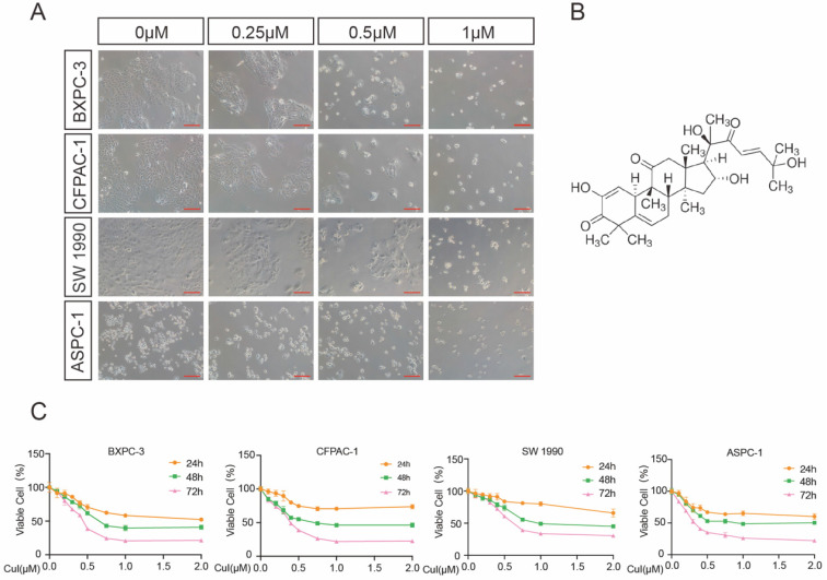
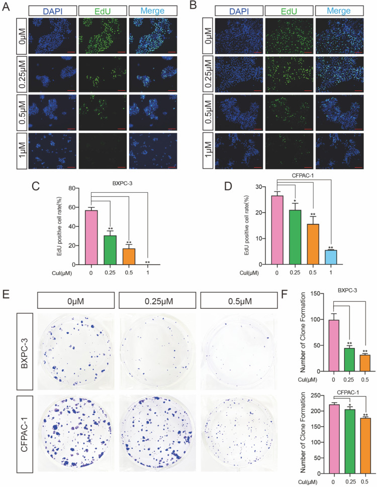
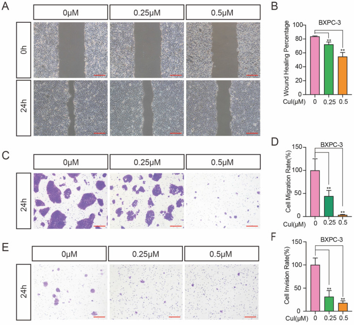
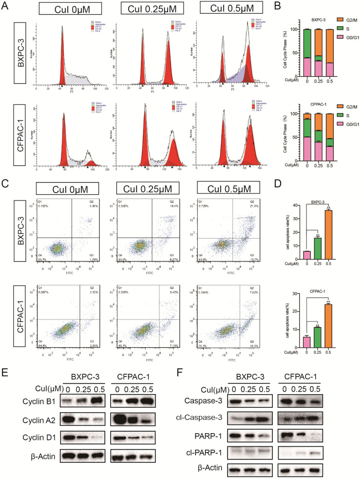
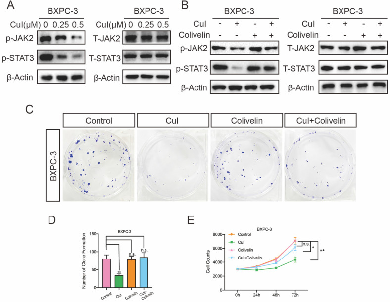
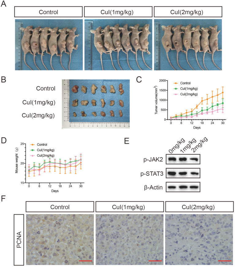
Pancreatic cancer is one of the most aggressive solid malignancies, as it has a 5-year survival rate of less than 10%. The growth and invasion of pancreatic cancer cells into normal tissues and organs make resection and treatment difficult. Finding an effective chemotherapy drug for this disease is crucial. In this study, we selected the tetracyclic triterpenoid compound cucurbitacin I, which may be used as a potential therapeutic drug for treating pancreatic cancer. First, we found that cucurbitacin I inhibited pancreatic cancer proliferation in a dose-time dependent manner. Further studies have shown that cucurbitacin I blocks the cell cycle of pancreatic cancer in the G2/M phase and induces cell apoptosis. In addition, under the action of the compound, the invasion ability of cells was greatly reduced and markedly impaired the growth of pancreatic tumour xenografts in nude mice. Furthermore, the decrease in pancreatic cancer cell proliferation caused by cucurbitacin I appeared to involve JAK2/STAT3 signalling pathway inhibition, and the use of JAK2/STAT3 activators effectively restored the inhibition. In conclusion, our research may provide a basis for the further development of pancreatic cancer treatment drugs.
Keywords: Cucurbitacin I; JAK2/STAT3 signalling pathway; pancreatic cancer; proliferation.
© The author(s).
Conflict of interest statement
Competing Interests: The authors have declared that no competing interest exists.
Figures
Similar articles
-
Cucurbitacin B inhibits cell proliferation and induces apoptosis in human osteosarcoma cells via modulation of the JAK2/STAT3 and MAPK pathways.Exp Ther Med. 2017 Jul;14(1):805-812. doi: 10.3892/etm.2017.4547. Epub 2017 Jun 6. Exp Ther Med. 2017. PMID: 28673003 Free PMC article.
-
Cucurbitacin B controls M2 macrophage polarization to suppresses metastasis via targeting JAK-2/STAT3 signalling pathway in colorectal cancer.J Ethnopharmacol. 2022 Apr 6;287:114915. doi: 10.1016/j.jep.2021.114915. Epub 2021 Dec 24. J Ethnopharmacol. 2022. PMID: 34954267
-
Anticancer effect of cucurbitacin B on MKN-45 cells via inhibition of the JAK2/STAT3 signaling pathway.Exp Ther Med. 2016 Oct;12(4):2709-2715. doi: 10.3892/etm.2016.3670. Epub 2016 Sep 6. Exp Ther Med. 2016. PMID: 27698776 Free PMC article.
-
STAT3-independent inhibition of lysophosphatidic acid-mediated upregulation of connective tissue growth factor (CTGF) by cucurbitacin I.Biochem Pharmacol. 2006 Jun 28;72(1):32-41. doi: 10.1016/j.bcp.2006.04.001. Epub 2006 Apr 25. Biochem Pharmacol. 2006. PMID: 16707113
-
Cucurbitacin B and SCH772984 exhibit synergistic anti-pancreatic cancer activities by suppressing EGFR, PI3K/Akt/mTOR, STAT3 and ERK signaling.Oncotarget. 2017 Oct 9;8(61):103167-103181. doi: 10.18632/oncotarget.21704. eCollection 2017 Nov 28. Oncotarget. 2017. PMID: 29262554 Free PMC article.
Cited by
-
Progranulin promoted the proliferation, metastasis, and suppressed apoptosis via JAK2-STAT3/4 signaling pathway in papillary thyroid carcinoma.Cancer Cell Int. 2023 Sep 2;23(1):191. doi: 10.1186/s12935-023-03033-2. Cancer Cell Int. 2023. PMID: 37660003 Free PMC article.
-
Synthesis and biological evaluation of thiourea-tethered benzodiazepinones as anti-proliferative agents targeting JAK-3 kinase.Naunyn Schmiedebergs Arch Pharmacol. 2025 Aug;398(8):10667-10689. doi: 10.1007/s00210-025-03853-1. Epub 2025 Mar 3. Naunyn Schmiedebergs Arch Pharmacol. 2025. PMID: 40029384
-
Cucurbitacins as Potent Chemo-Preventive Agents: Mechanistic Insight and Recent Trends.Biomolecules. 2022 Dec 27;13(1):57. doi: 10.3390/biom13010057. Biomolecules. 2022. PMID: 36671442 Free PMC article. Review.
-
Cucurbitacin I Reverses Tumor-Associated Macrophage Polarization to Affect Cancer Cell Metastasis.Int J Mol Sci. 2023 Nov 2;24(21):15920. doi: 10.3390/ijms242115920. Int J Mol Sci. 2023. PMID: 37958903 Free PMC article.
-
Recent Advances in the Application of Cucurbitacins as Anticancer Agents.Metabolites. 2023 Oct 14;13(10):1081. doi: 10.3390/metabo13101081. Metabolites. 2023. PMID: 37887406 Free PMC article. Review.
References
-
- Siegel RL, Miller KD, Fuchs HE, Jemal A. Cancer Statistics, 2021. CA Cancer J Clin. 2021;71:7–33. - PubMed
-
- Sung H, Ferlay J, Siegel RL, Laversanne M, Soerjomataram I, Jemal A. et al. Global Cancer Statistics 2020: GLOBOCAN Estimates of Incidence and Mortality Worldwide for 36 Cancers in 185 Countries. CA Cancer J Clin. 2021;71:209–49. - PubMed
-
- Kindler HL. A Glimmer of Hope for Pancreatic Cancer. N Engl J Med. 2018;379:2463–4. - PubMed
-
- Kim MP, Gallick GE. Gemcitabine resistance in pancreatic cancer: picking the key players. Clin Cancer Res. 2008;14:1284–5. - PubMed
LinkOut - more resources
Full Text Sources
Miscellaneous
