. 2022 Apr 19:10:877047.
doi: 10.3389/fcell.2022.877047. eCollection 2022.

Canonical Wnt Signaling Promotes Formation of Somatic Permeability Barrier for Proper Germ Cell Differentiation

Affiliations

Canonical Wnt Signaling Promotes Formation of Somatic Permeability Barrier for Proper Germ Cell Differentiation

Ting-An Chen et al. Front Cell Dev Biol. .

Abstract

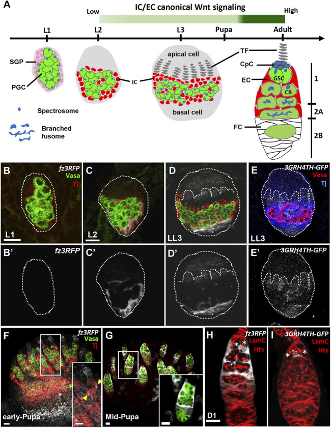
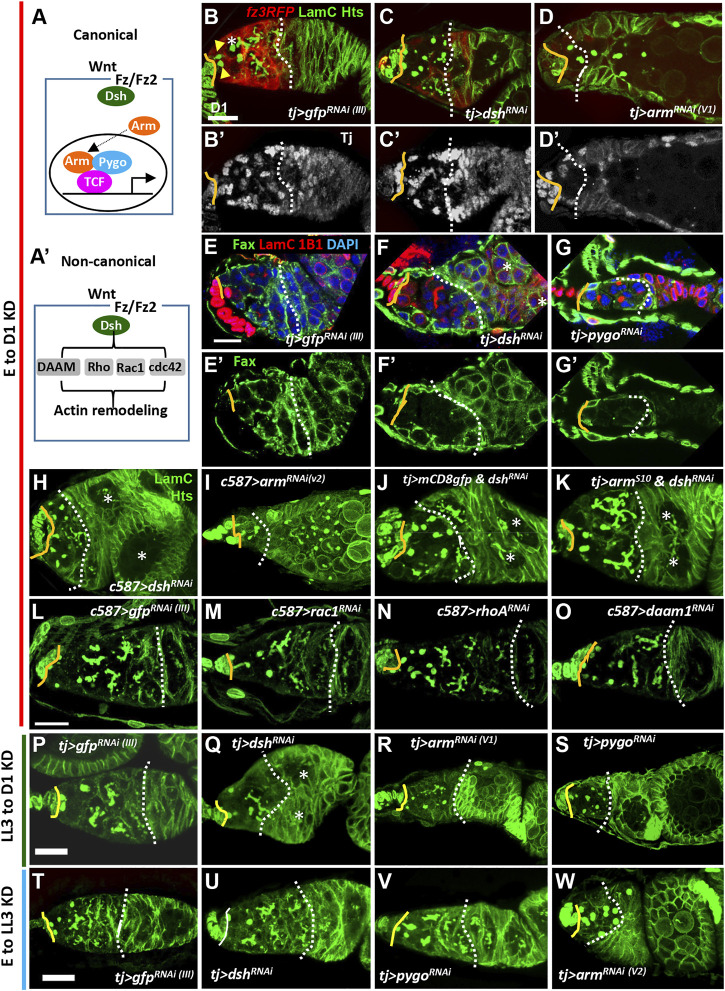
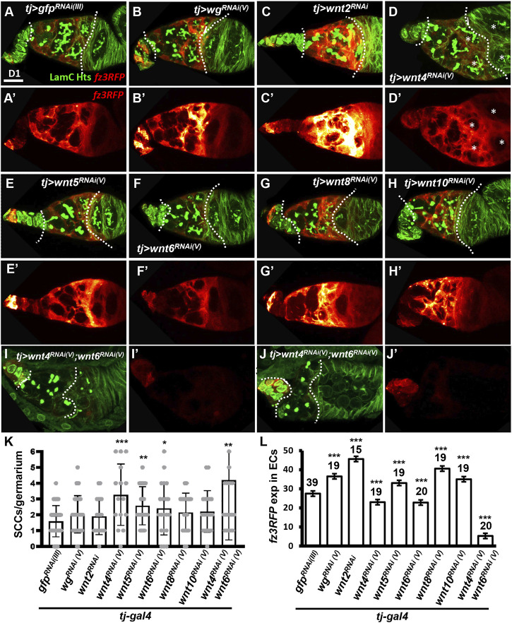
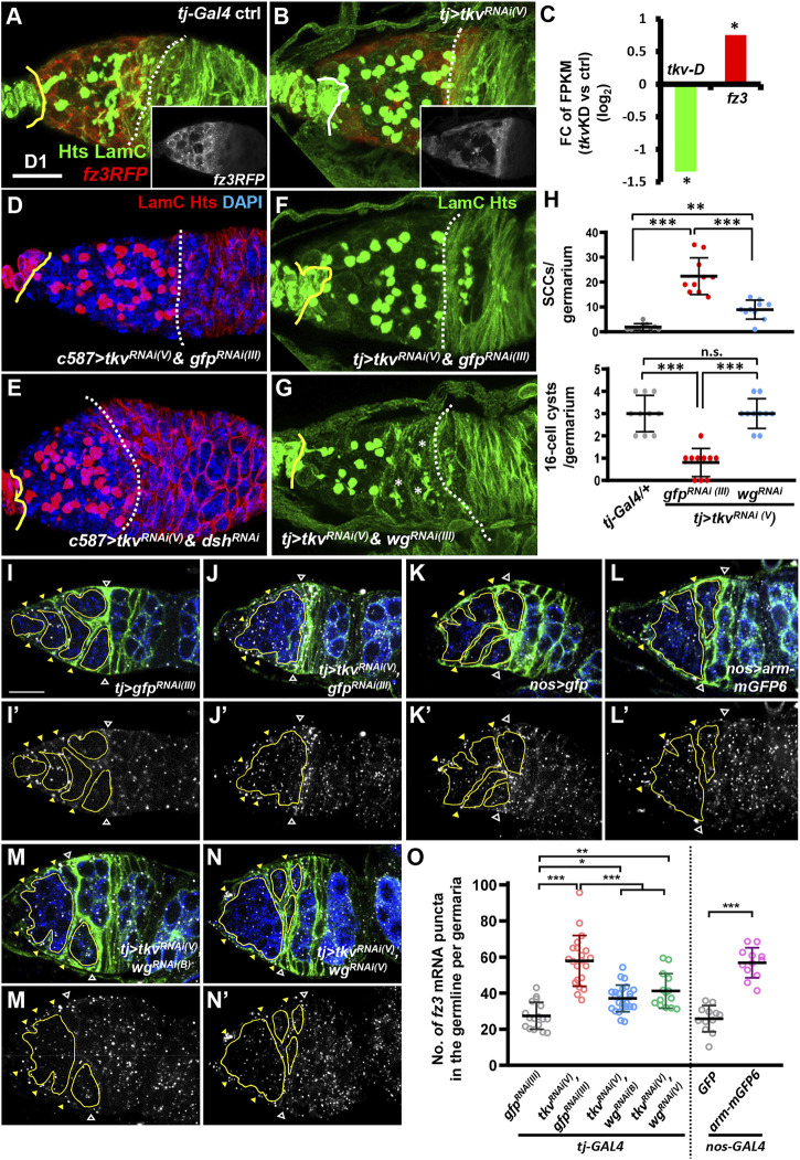
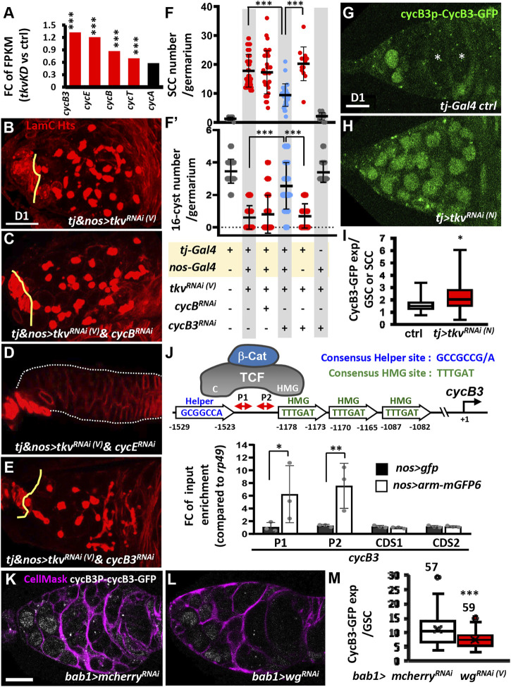
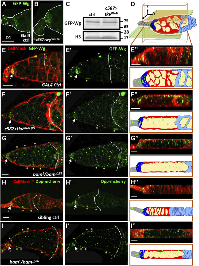
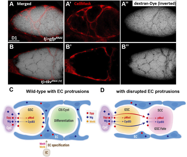
Morphogen-mediated signaling is critical for proper organ development and stem cell function, and well-characterized mechanisms spatiotemporally limit the expression of ligands, receptors, and ligand-binding cell-surface glypicans. Here, we show that in the developing Drosophila ovary, canonical Wnt signaling promotes the formation of somatic escort cells (ECs) and their protrusions, which establish a physical permeability barrier to define morphogen territories for proper germ cell differentiation. The protrusions shield germ cells from Dpp and Wingless morphogens produced by the germline stem cell (GSC) niche and normally only received by GSCs. Genetic disruption of EC protrusions allows GSC progeny to also receive Dpp and Wingless, which subsequently disrupt germ cell differentiation. Our results reveal a role for canonical Wnt signaling in specifying the ovarian somatic cells necessary for germ cell differentiation. Additionally, we demonstrate the morphogen-limiting function of this physical permeability barrier, which may be a common mechanism in other organs across species.

Keywords: DPP; GSCs; TKV; Wg; escort cell protrusions; germline stem cells.


Conflict of interest statement

The authors declare that the research was conducted in the absence of any commercial or financial relationships that could be construed as a potential conflict of interest.

Figures

FIGURE 1
FIGURE 1
Canonical Wnt signaling is activated in the somatic cells of developing ovaries. (A) Schematic of Drosophila larval gonads and the adult germarium. Primordial germ cells (PGCs), each with a round-shaped fusome (unique membrane-enriched organelle), and somatic gonad precursors (SGPs) are present at the L1 and L2 stages. PGC and SGP numbers are greatly increased at the L3 stage, and SGPs differentiate into apical cells, terminal filaments (TFs), intermingled cells (ICs) and basal cells. ICs later further differentiate into cap cells (CpCs), escort cells (ECs), and probably follicle cells (FCs). During the pupal stage, apical cells migrate through TFs, ICs and basal cells to generate ovarioles; the anterior structure of the ovariole is the germarium. In the adult germarium, GSCs and their progeny are wrapped by EC protrusions in regions 1 and 2A. Then, ECs are replaced by follicle cells (FCs) in the 2B region. During the growth of GSC progeny, fusomes become branched. The green bar indicates canonical Wnt signaling is detectable in the EC precursors of LL3 animals, and it becomes strongly activated in ECs at the mid-pupa stage, persisting into the adult fly. (BI) L1 (B), L2 (C) and late-L3 gonads (D and E) with fz3RFP in B-D (gray, canonical Wnt signaling reporter), 3GRH4TH-GFP in E (gray, canonical Wnt signaling reporter), Vasa (green in B–D, red in E, PGCs) and Tj (red in B-D, blue in E, ICs). (B’–D’) and E only show fz3RFP and 3GRH4TH-GFP channel, respectively. Dashed circles outline the gonad; dashed line marks a forming TF. (FI) Early-pupa (F), Mid-pupa (G) and adult day 1 germaria (H and I) with fz3RFP in F-H (gray, canonical Wnt signaling reporter), 3GRH4TH-GFP in I (gray, canonical Wnt signaling reporter), Vasa (green in F and G, germ cells), Tj (red in F and G, EC and follicle cell nuclei), LamC (red in H and I, TF and cap cell nuclear envelopes), and Hts (red in H and I, fusomes). Insets in F and G show enlarged images from the corresponding dashed squares in F and G. Arrowheads in F inset show ECs with fz3RFP. The genotype in F is nos > gfp RNAi . Asterisks in H and I mark GSCs. Scale bars are 10 μm.
FIGURE 2
FIGURE 2
Canonical Wnt signaling in the developing soma controls escort cell formation and promotes germ cell differentiation. (A and A’) Canonical (A) and non-canonical Wnt signaling pathways (A’). Wnt signaling components were knocked down in the ovarian soma from embryo to adult day (D)1 (B–O), from late-L3 (LL3) to adult D1 (P–S), and from embryo to LL3 (T–W), and D1 germaria were examined. (B–O) tj-GAL4>gfp RNAi (III) (B and E) , tj > dsh RNAi (C and F), tj > arm RNAi (V1) (D), tj > pygo RNAi (V) (G), c587>dsh RNAi (H), and c587>arm RNAi(V) (I), tj > mCD8-gfp & dsh RNAi (J), tj > arm S10 & dsh RNAi (K), c587>gfp RNAi (III) (L), c587>rac RNAi (M), c587>rhoA RNAi (N) and c587>daam1 RNAi germaria (O) with LamC (green in B–D and H–O, red in E–G, terminal filament (TF) and cap cell nuclear envelopes), Hts (green in B–D and H–O, red in E–G, fusomes), Tj in (B’ to D’) (gray, nuclei of cap, escort and follicle cells), Fax (green in E-G, EC membranes), and DAPI (blue in E-G, DNA). (E’–G’) show only Fax channel. (P–S) tj-GAL4>gfp RNAi (III) (P), tj > dsh RNAi (Q), tj > arm RNAi (V1) (R) and tj > pygo RNAi germaria (S) with LamC (green) and Hts (green). (T–W) tj-GAL4>gfp RNAi (III) (T), tj > dsh RNAi (U), tj > pygo RNAi (N) and tj > arm RNAi (V2) germaria (W) with LamC (green) and Hts (green). Solid lines, GSC-cap cell junction; dashed lines, the 2A/2B boundary (or the junction between escort cells and follicle cells when 2A/2B boundary is missing). Arrowheads and an asterisk in panel 2B respectively mark GSCs and cystoblast. Two asterisks in F, H, K, and Q denote two side-by-side egg chambers in the ovariole. Scale bars are 10 μm; B-D and H–K, E–G, L–O, P–S, and T–W have the same scale bar.
FIGURE 3
FIGURE 3
Wnt4 and Wnt6 activate canonical Wnt signaling in escort cells. (A–H) One-day (D)-old tj > gfp RNAi (III) (A), tj > wg RNAi(V) (B), tj > wnt2 RNAi (C), tj > wnt4 RNAi(V) (D), tj > wnt5 RNAi(V) (E), tj > wnt6 RNAi(V) (F), tj > wnt8 RNAi(V) (G), tj > wnt10 RNAi(V) germaria (H) and tj > wnt4 RNAi(V) &wnt6 RNAi(V) ( (I and J) with fz3RFP (canonical Wnt signaling reporter), LamC (green, terminal filament and cap cell nuclear envelopes), and Hts (green, fusomes). (A’–J’), Heatmap showing relative intensity, red = low to yellow = high, of fz3RFP in the germaria. Images were processed with Zen blue and Fire look-up table (LUT); color gradient bar indicates strength of fz3RFP from low (L, red) to High (L, gold). Scale bar, 10 μm; A–H have the same scale bar, K and L have the same scale bar. (K) Number of spectrosome-containing cells (SCCs) per germarium for indicated genotypes. (L) Expression of fz3RFP in the escort cell region (between cap cells and follicle cells; marked by dashed lines) of germaria with the indicated genotypes. RNAi was expressed throughout development until dissection. Error bar, mean ± SD. Statistical analysis, One-way ANOVA, *, p < 0.05; **, p < 0.01; ***, p < 0.001.
FIGURE 4
FIGURE 4
Wg signaling is activated in the germ cells of somatic-tkv knockdown germaria. (A and B) tj-GAL4/+ (A) and tj > tkv RNAi (V) germaria (B) with LamC (green, TF and cap cell nuclear envelopes), Hts (green, fusomes) and fz3RLFP (red, canonical Wnt signaling reporter). Insets in A and B show only fz3RFP channel. (C) Fold-change (FC) of RNA-seq based gene expression values (log2) for tkv transcript variant D (tkv-D) and fz3 in 1-day (D)-old control (ctrl, UAS-tkv RNAi (N) /+) and c587>tkv RNAi (N) anterior ovarioles compared with UAS-tkv RNAi (N) /+ (control, ctrl). FPKM, fragments per kilobase of transcript per million mapped reads. *, p < 0.05. Statistical analysis was performed with two biological replicates. (D to G) c587>tkv RNAi (V) & gfp RNAi (III) (D), c587>tkv RNAi (V) & dsh RNAi (E), tj > tkv RNAi (V) & gfp RNAi (III) (F) and c587>tkv RNAi (V) & wg RNAi germaria (G) with LamC (red in D and E, green in F and G), Hts (red in D and E, green in F and G) and DAPI (blue, DNA, in D and E). Yellow lines denote the junction between cap cell and GSC; dashed line mark the 2A/2B boundary or the junction between escort cells and follicles when the 2A/B boundary is missing. Asterisks mark 16-cell cysts. (H) Numbers of spectrosome-containing cells (SCCs) and 16-cell cysts per germarium of flies with the indicated genotypes. (I–N) In situ hybridized tj > gfp RNAi (III) (I), tj > tkv RNAi (V) & gfp RNAi (III) (J), tj > gfp (K), tj > arm-mGFP6 (L), tj > tkv RNAi (V) & wg RNAi(V) (M) and tj > tkv RNAi (V) & wg RNAi(B) germaria (N) with labeling for Fax (green, escort cell membrane extension), Vasa-GFP (blue, germ cells), and fz3 mRNA (gray). (I’N’) show the fz3 channel. Hollow triangles point to the 2A/B boundary; yellow triangles indicate escort cell region; germ cell regions before the 2A/B boundary are outlined by yellow circles. (O) Number (No.) of fz3 mRNA puncta in the germline per germarium with the indicated genotypes. *, p < 0.05, **, p < 0.01; ***, p < 0.001. Error bars indicate mean ± S.D., One-Way ANOVA was used for statistical analysis. RNAi was expressed throughout development. Scale bar, 10 μm. A and B, D-G, and I to N are 3D-reconstructed images.
FIGURE 5
FIGURE 5
Canonical Wnt signaling transcriptionally activates cycB3 to suppress differentiation in the germline of somatic-tkv knockdown germaria. (A) Fold-change (FC) of RNA-seq-based gene expression values (log2) for indicated cyclin (cyc) gene in 1-day (D)-old control (ctrl, UAS-tkv RNAi(N) /+) and c587>tkv RNAi(N) anterior ovarioles compared with UAS-tkv RNAi(N) /+ (control, ctrl). FPKM, fragments per kilobase of transcript per million mapped reads. ***, p < 0.001. Statistical analysis was performed with two biological replicates. (B to E) One-day-old tj & nos > tkv RNAi (V) () (B), tj & nos > tkv RNAi(V) & cycB RNAi (C), tj & nos > tkv RNAi(V) & cycE RNAi (D) and tj & nos > tkv RNAi (V) & cycB3 RNAi (E) and with LamC (red, terminal filament and cap cell nuclear envelopes) and Hts (red, fusomes). Solid lines mark junction between GSCs and cap cells; dashed lines outline the germaria in D. (F and F’) Numbers of spectrosome-containing cells (SCCs) (F), and 16-cell cysts per germarium (F’) of flies with the indicated genotypes. (G and H) Live image of 1-day-old tj > GFP RNA (III) (G) and tj > tkv RNAi (V) germaria (H) bearing cycB3P-cycB3-gfp (Green, CycB3-GFP). (I) Box plot shows expression of CycB3-GFP in GSCs or SCCs in the indicated genotypes. (J) Schematic shows how β-catenin (β-Cat) interacts with TCF, which binds to HMG and Helper sites of the cycB3 promoter through its HMG and C domains, respectively. ChIP analysis of TCF binding in 1-day-old ovaries; the chromatin from nos > gfp and nos > arm-mgfp6 cells was precipitated with GFP-Trap beads. Co-precipitated DNA was analyzed by qPCR using two sets of primers (P1 and P2) against the region between Helper and HMG sites. The amplicons of two different coding regions were used as negative controls. (K and L) One-day-old bab1>mcherry RNAi (K) and bab1>wg RNAi (V) (L) with cycB3P-CycB3-GFP (gray) and CellMask (Magenta, cell membrane). (M) Average of CycB3-GFP expression in GSCs of indicated genotypes. Number of GSCs analyzed is shown above each bar. Differences in F and F′ were analyzed by one-way ANOVA; data in I and M were analyzed by Student’s t test, and in J were analyzed by two-way ANOVA. Solid line in the box of I and M is median; cross in M is Mean. Error bars represent SD; *, p < 0.05; **, p < 0.01; ***, p < 0.001. RNAi was expressed throughout development until dissection. Scale bar is 10 μm.
FIGURE 6
FIGURE 6
Cellular protrusions of escort cells serve as a physical permeability barrier to prevent Wg and Dpp distribution in the germline. (A and B) Live images of anterior part of 1-day (D)-old GAL4 control (ctrl) (A) and c587>wg RNAi germaria (B) bearing GFP-wg (green). Dashed lines show the edge of the germarium. (C) Representative immunoblot shows that Wg-GFP expression (anti-GFP antibody) is similar in 1-day (D)-old control (ctrl) and c587>tkv RNAi (V) ovaries. Histone (H3) was used as a loading control. Molecular weight markers are indicated to the right of the blots. (D) Schematic of a germarium with a three-axis (X, Y, and Z) coordinate system. The directions of each axis are shown. The Y-axis is defined as anterior to posterior. An XY section is shown as the light green shaded area, and an XZ section is shown as a light pink shaded area. Terminal filament cells, gray; cap cells, dark green; escort cells, red; germ cell, light yellow; follicle cells, light blue. (E–I) Live images of 1-day-old GAL4 control (E), c587>tkv RNAi(v) (F), and bam 1 /bam △86 mutant (G and I), sibling control germaria (H), bearing GFP-wg (green in E-G), Dpp-mcherry (green in H and I) and labelled with CellMask (red, cell membrane). (E”–I”) are optical sections in the XZ plane; the corresponding schematic with the cell types is shown in D and corresponds to (E’–I’); green color shows the distribution of Wg (E”–G”) or Dpp (H”–I”). Scale bar, 10 μm; A and B, E and F, and H and I share the same scale bar. Germaria were examined for wg-GFP distribution in control (n = 12), c587>tkv RNAi(v) (n = 25), and bam 1 /bam △86 mutant (n = 15). Germaria were examined for Dpp-mechrry distribution in the control (n = 8), and bam 1 /bam △86 mutant (n = 12). Control genotypes in A, C and E are c587>gfp RNAi(III) , GFP-wg/+; in H is (+/Dpp-mcherry; bam 1 or bam △86 /+). Arrows point to the cap cell region. Dashed lines in E and H mark the 2A/2B boundary, and F, G and H mark in the junction between escort cells and follicle cells (the 2A/2B boundary is lost). Asterisks mark GFP signals present in ECs.
FIGURE 7
FIGURE 7
A permeability assay for the ovary assay reveals the role of EC protrusions warpping germ cells and acting as a physical permeability barrier. (A and B) One-day-old live tj > gfp RNAi (A) and tj > tkv RNAi(V) germaria (B) with CellMask (red, EC cell membrane) and dextran-488 (dextra signals are inverted for better visualization, a fluorescence dye). A and B are merged images; (A’ and B’) show only CellMask channer and (A” and B”) show only dextran-488 channel. Scale bar, 10 μm. (C and D) Model of canonical Wnt signaling in EC specification and promotion of EC protrusions to set Dpp and Wg territories and maintain germline homeostasis. (C) In the developing wildtype ovary, canonical Wnt signaling in intermingled cells (ICs) is at least in part activated by Wnt4 and Wnt6. This signaling is critical for escort cell (EC) formation and maintains EC protrusions. The EC protrusions serve to compartmentalize GSC progeny and shield the germ cells from Dpp and Wg produced by cap cells (CpC), allowing the GSC progeny to properly differentiate. In GSCs, Dpp signaling leads to Mad phosphorylation (pMad), and upregulation of CycB3 probably occurs via transcriptional activation by Wg signaling. These events are critical to maintain GSC fate. (D) In the germarium with blunted EC protrusions, Dpp and Wg also signal to GSC progeny and disrupt their differentiation. CB, cytoblast; cyst, germ cell cysts.

Similar articles

Cited by

References

    1. Ables E. T., Drummond-Barbosa D. (2013). Cyclin E controlsDrosophilafemale Germline Stem Cell Maintenance Independently of its Role in Proliferation by Modulating Responsiveness to Niche Signals. Development (Cambridge, England) 140, 530–540. 10.1242/dev.088583 - DOI - PMC - PubMed
    1. Aradhya R., Zmojdzian M., Da Ponte J. P., Jagla K. (2015). Muscle Niche-Driven Insulin-Notch-Myc cascade Reactivates Dormant Adult Muscle Precursors in Drosophila. Elife 4. 10.7554/eLife.08497 - DOI - PMC - PubMed
    1. Aramaki S., Hayashi K., Kurimoto K., Ohta H., Yabuta Y., Iwanari H., et al. (2013). A Mesodermal Factor, T, Specifies Mouse Germ Cell Fate by Directly Activating Germline Determinants. Developmental Cel 27, 516–529. 10.1016/j.devcel.2013.11.001 - DOI - PubMed
    1. Ashburner M. (2005). Drosophila: A Laboratory Handbook. NY: Cold Spring Harbor Laboratory Press, Cold Spring Harbor.
    1. Banisch T. U., Maimon I., Dadosh T., Gilboa L. (2017). Escort Cells Generate a Dynamic Compartment for Germline Stem Cell Differentiation via Combined Stat and Erk Signalling. Development 144, 1937–1947. 10.1242/dev.143727 - DOI - PubMed

LinkOut - more resources