Review
. 2022 Apr 20:13:825266.
doi: 10.3389/fphar.2022.825266. eCollection 2022.

Oral Potentially Malignant Disorders: Etiology, Pathogenesis, and Transformation Into Oral Cancer

Affiliations
Review

Oral Potentially Malignant Disorders: Etiology, Pathogenesis, and Transformation Into Oral Cancer

Pratima Kumari et al. Front Pharmacol. .

Abstract

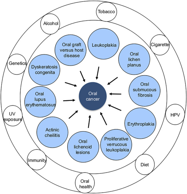
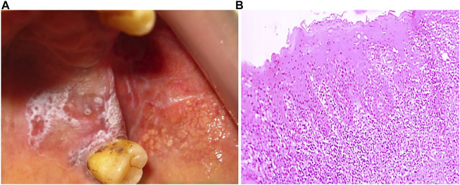
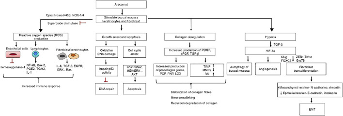
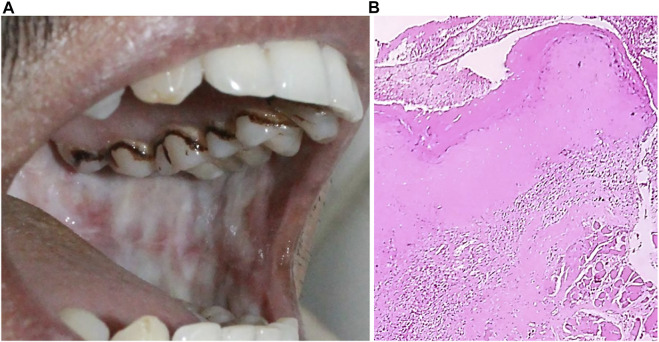
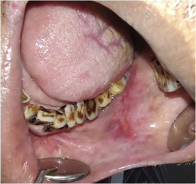
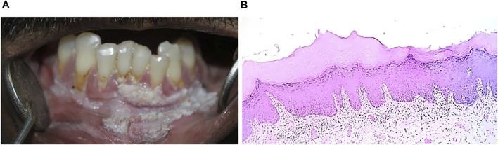
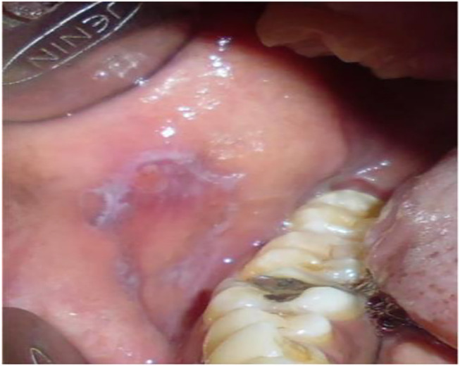
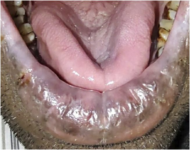
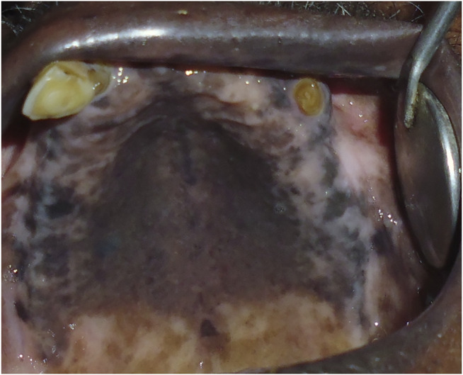
Among oral diseases, oral cancer is a critical health issue due to its life-threatening potential. Globocan, in its 2020 report, estimated ∼0.37 million new cases of oral cancer, with the majority of them coming from the Asian continent. The WHO has anticipated a rise in the incidences of oral cancer in the coming decades. Various factors, such as genetic, epigenetic, microbial, habitual, and lifestyle factors, are closely associated with oral cancer occurrence and progression. Oral lesions, inherited genetic mutations (dyskeratosis congenital syndrome), and viral infections (HPV) are early signs of oral cancer. Lesions with dysplastic features have been categorized under oral potentially malignant disorders (OPMDs), such as oral leukoplakia, erythroplakia, oral submucous fibrosis (OSMF), and proliferative verrucous leukoplakia, are assumed to have a high risk of malignancy. The incidence and prevalence of OPMDs are recorded as being high in South-Asian countries. Early detection, prevention, and treatment of OPMDs are needed to prevent its malignant transformation into oral cancer. Many advanced diagnostic techniques are used to predict their progression and to assess the risk of malignant transformation. This communication provides insight into the importance of early detection and prevention of OPMDs.

Keywords: erythroplakia; leukoplakia; malignant transformation; oral lichen planus; oral lichenoid lesions; oral potentially malignant disorders; oral submucous fibrosis; proliferative verrucous leukoplakia.


Conflict of interest statement

The authors declare that the research was conducted in the absence of any commercial or financial relationships that could be construed as a potential conflict of interest.

Figures

FIGURE 1
FIGURE 1
Systematic representation of normal buccal mucosa.
FIGURE 2
FIGURE 2
Different types of OPMDs (inner circle) and common risk factors (outer rings) associated with oral cancer development.
FIGURE 3
FIGURE 3
(A) Clinical presentation of leukoplakia, a typical appearance of white patch. (B) H&E stained section (4x) showing hyperkeratinized stratified squamous epithelium.
FIGURE 4
FIGURE 4
Role of immune cells in OLP pathogenesis; (A) Immune infiltration in OLP, CD8+ T cells mediated hyperimmune activation leading to apoptosis of basal keratinocytes. (B) Possible mechanism of hyperimmune response in buccal mucosa, where CD8+ T cells are activated by oral keratinocytes and CD4+ T cells.
FIGURE 5
FIGURE 5
(A) Oral lichen planus (B) Hyperkeratinized stratified squamous epithelium (10x) showing basal cell degeneration at focal areas. Juxtaepithelially stroma shows presence of lymphocytes infiltration.
FIGURE 6
FIGURE 6
Hyperkeratotic epithelial cells in OSMF. Immune activation in OSMF show the presence of different types of immune infiltrates in submucous layer. Increase in fibroblast cells leads to high production of collagen fibres (brown bar) leading to fibrosis of oral mucosa.
FIGURE 7
FIGURE 7
OSMF pathogenesis: Cellular and molecular changes in buccal mucosa due to areca nut exposure.
FIGURE 8
FIGURE 8
(A) Oral submucosal fibrosis of cheek mucosa. (B) Keratinized stratified squamous epithelium (4x) showing presence of hyalinization and collagenous connective tissue stroma.
FIGURE 9
FIGURE 9
Erythroplakia patch.
FIGURE 10
FIGURE 10
(A) Proliferative verrucous leukoplakia show multifocal presence in anterior gingiva, labial vestibule extended to right and left side of gingiva. (B) Hyperkeratinized stratified squamous epithelium (4x) with verrucous like projection and inflammatory cell infiltration in lamina propria.
FIGURE 11
FIGURE 11
Clinical representation of oral lichenoid lesions.
FIGURE 12
FIGURE 12
Actinic cheilitis a typically presentation at lower lip.
FIGURE 13
FIGURE 13
Clinical presentation of palatal lesions in reverse smokers.

Similar articles

Cited by

References

    1. Abadie W. M., Partington E. J., Fowler C. B., Schmalbach C. E. (2015). Optimal Management of Proliferative Verrucous Leukoplakia: a Systematic Review of the Literature. Otolaryngol. Head Neck Surg. 153 (4), 504–511. 10.1177/0194599815586779 - DOI - PubMed
    1. Abidullah M., G K. K., Mawardi H., Alyami Y., Arif S. M., Qureshi Y. (2018). Clinical and Histopathological Correlation of Oral Submucous Fibrosis- an Institutional Study. jemds 7 (18), 2227–2230. 10.14260/jemds/2018/501 - DOI
    1. Al-Swiahb J. N., Chen C. H., Chuang H. C., Fang F. M., Tasi H. T., Chien C. Y. (2010). Clinical, Pathological and Molecular Determinants in Squamous Cell Carcinoma of the Oral Cavity. Future Oncol. 6 (5), 837–850. 10.2217/fon.10.35 - DOI - PubMed
    1. Amin N. R., Yussif N., Ahmed E. (2020). The Effect of Smoking on Clinical Presentation and Expression of TLR-2 and CD34 in Oral Lichen Planus Patients: Clinical and Immunohistochemical Study. BMC oral health 20, 129–135. 10.1186/s12903-020-01118-2 - DOI - PMC - PubMed
    1. Angadi P. V., Rao S. S. (2011). Areca Nut in Pathogenesis of Oral Submucous Fibrosis: Revisited. Oral Maxillofac. Surg. 15 (1), 1–9. 10.1007/s10006-010-0219-8 - DOI - PubMed

LinkOut - more resources