. 2022 Apr 19:9:871499.
doi: 10.3389/fmolb.2022.871499. eCollection 2022.

Targeting the N-Terminus Domain of the Coronavirus Nucleocapsid Protein Induces Abnormal Oligomerization via Allosteric Modulation

Affiliations

Targeting the N-Terminus Domain of the Coronavirus Nucleocapsid Protein Induces Abnormal Oligomerization via Allosteric Modulation

Jia-Ning Hsu et al. Front Mol Biosci. .

Erratum in

Abstract

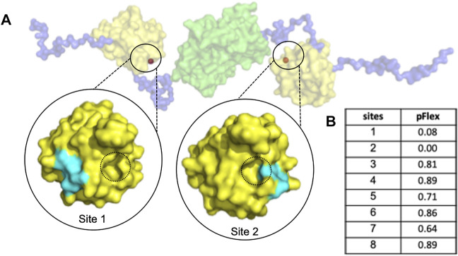
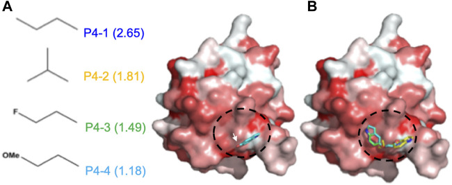
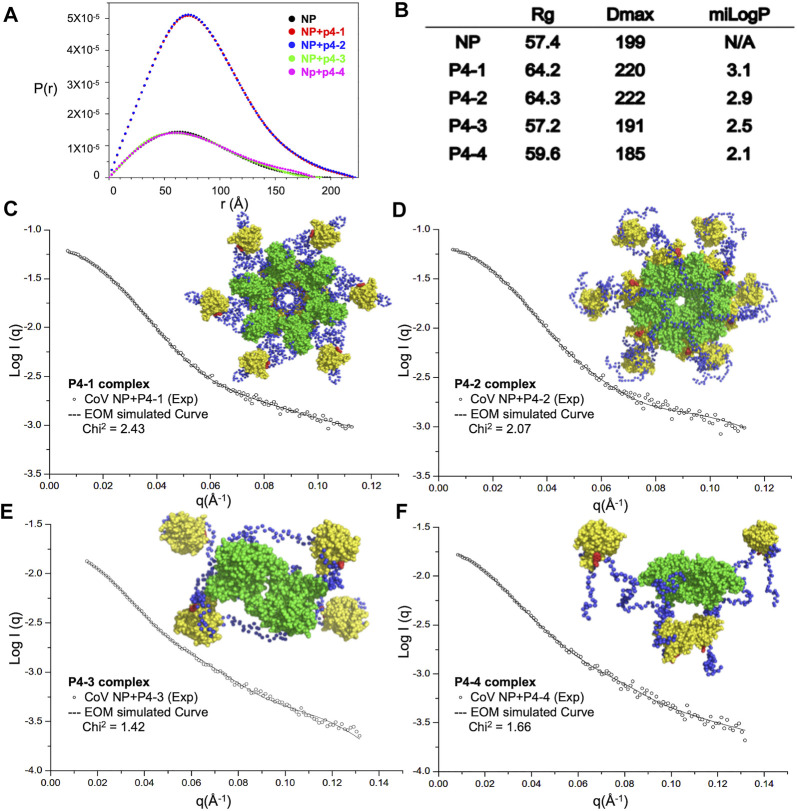
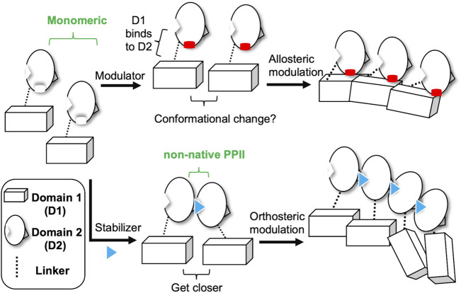
Epidemics caused by coronaviruses (CoVs), namely the severe acute respiratory syndrome (SARS) (2003), Middle East respiratory syndrome (MERS) (2012), and coronavirus disease 2019 (COVID-19) (2019), have triggered a global public health emergency. Drug development against CoVs is inherently arduous. The nucleocapsid (N) protein forms an oligomer and facilitates binding with the viral RNA genome, which is critical in the life cycle of the virus. In the current study, we found a potential allosteric site (Site 1) using PARS, an online allosteric site predictor, in the CoV N-N-terminal RNA-binding domain (NTD) to modulate the N protein conformation. We identified 5-hydroxyindole as the lead via molecular docking to target Site 1. We designed and synthesized four 5-hydroxyindole derivatives, named P4-1 to P4-4, based on the pose of 5-hydroxyindole in the docking model complex. Small-angle X-ray scattering (SAXS) data indicate that two 5-hydroxyindole compounds with higher hydrophobic R-groups mediate the binding between N-NTD and N-C-terminal dimerization domain (CTD) and elicit high-order oligomerization of the whole N protein. Furthermore, the crystal structures suggested that these two compounds act on this novel cavity and create a flat surface with higher hydrophobicity, which may mediate the interaction between N-NTD and N-CTD. Taken together, we discovered an allosteric binding pocket targeting small molecules that induces abnormal aggregation of the CoV N protein. These novel concepts will facilitate protein-protein interaction (PPI)-based drug design against various CoVs.

Keywords: COVID-19; MERS-CoV; PPI-based drug design; allosteric modulator; n protein.


Conflict of interest statement

The authors declare that the research was conducted in the absence of any commercial or financial relationships that could be construed as a potential conflict of interest.

Figures

FIGURE 1
FIGURE 1
The dimeric MERS-CoV N protein possesses a druggable sites for allosteric modulator design. (A) The structure of dimeric N protein was obtained by SAXS experiment from previous publication (Lin et al., 2020) in which the NTD, linker and dimeric CTD were shown in yellow, blue and green, respectively. The predicted allosteric sites were shown in orange and highlighted in black circles. For simplification, only the sites with potent pFlex were shown. The interface of non-native PPI was indicated with black dotted circles. (B) The pFlex values of all predicted sites. pFlex indicates overall flexibility of target protein may be affected by the binding ligand to the sites where the p-value is lower than 0.05.
FIGURE 2
FIGURE 2
Docking results of CoV N-NTD with 5-Hydroxyindole. (A) (left) The structures of each chemical moiety designed for 5-Hydroxyindole modification. The calculated miLogP values of each moiety were shown in brackets. (right) Surface representation of CoV N-NTD with the expected binding site of 5-Hydroxyindole, obtained by using the molecular docking. The surface was colored according to the hydrophobicity level at the protein surface. The chemical moieties were designed to add to the hydroxyl group of 5-Hydroxyindole (indicated by white arrow) to increase the hydrophobic contacts between each compound and the expected binding surface (indicated by black cycle) (B) Same as (A) except the 5-Hydroxyindole was replaced by P4-1, P4-2, P4-3, and P4-4 are shown in blue, yellow, green and cyan, respectively.
Scheme 1
Scheme 1
Synthesis of P4-1 to P4-4. Reagent and conditions: (A) NaH, DMF, 25°C, 30 min; then 1-iodopropane, 25°C, 3 h; (B) 2-bromopropane, NaH, DMF, 60°C, 6 h then 2-bromopropane, NaH, 60°C, 3h; (C) 1-bromo-2-fluoroethane, K2CO3, acetone, reflux, 12 h; (D) 1-iodo-2-methoxyethane, NaH, DMF, 25°C, 3 h.
FIGURE 3
FIGURE 3
Increased hydrophobicity of P4 ligand is correlated with the aggregation tendency of CoV N protein. (A) SAXS analysis of full-length CoV N protein complexed with P4 derivative compounds. Normalized results from GNOM are described with pairwise distance distribution P(r) and maximum distance. (B) The calculated values of Rg, Dmax and miLogP of each complex. (C–F) (left) Scattering profiles of P4-1 complex (C), P4-2 complex (D), P4-3 complex (E), and P4-4 complex (F) and normalization fitting with GNOM (dashed lines). (Right) Representative models of P4-1 complex (C), P4-2 complex (D), P4-3 complex (E), and P4-4 complex (F) generated by CRYSOL simulations of SAXS data. NTD, CTD, linker and P4s are shown as yellow, green, blue and red, respectively.
FIGURE 4
FIGURE 4
The detailed interactions between CoV N-NTD and each compound at P4-binding site. (Left) The CoV N protein was shown in cartoon and the residues involved in ligand binding were labeled and showed as sticks. (Right) Ligplot diagram of the interactions between N-NTD and P4 compounds. The contacting residues were labelled. Hydrophobic contacts and hydrogen bonds were displayed as red and green dashed lines, respectively. (A) The interactions of P4-1 complex. (B) The interactions of P4-2 complex. (C) The interactions of P4-3 complex. (D) The interactions of P4-4 complex.
FIGURE 5
FIGURE 5
Two different mechanisms of small-molecules targeting CoV N-NTD for inducing abnormal oligomerization. Allosteric modulator binds to the CoV N-NTD to create a hydrophobic flat surface that effect the oligomeric tendency of N-CTD, which induces the abnormal oligomerization of N protein. Whereas, N-NTD stabilizer acts on the non-native interface of CoV N-NTD dimer, which reduces the distance between targeted N proteins and eventually results in the aggregation of whole N protein.
FIGURE 6
FIGURE 6
Structural comparison of MERS CoV N-NTD with that of SARS-CoV-2 at P4 binding surface. (A) The structure of P4-1:MERS CoV N-NTD complex (yellow) was aligned to SARS-CoV-2 N-NTD (PDB: 6M3M), the interacting residues are highlighted in right box. (B) Protein sequence alignment of N-NTD of various CoVs. The conservation scoring was calculated by PRALINE and indicated as 0 (least conserved) to 10 (most conserved). The residues involved in P4-1 binding were indicated by black box.

Similar articles

Cited by

References

    1. Adams P. D., Grosse-Kunstleve R. W., Hung L.-W., Ioerger T. R., McCoy A. J., Moriarty N. W., et al. (2002). PHENIX: Building New Software for Automated Crystallographic Structure Determination. Acta Crystallogr. D Biol. Cryst. 58 (Pt 11), 1948–1954. 10.1107/s0907444902016657 - DOI - PubMed
    1. Almazán F., Galán C., Enjuanes L. (2004). The Nucleoprotein Is Required for Efficient Coronavirus Genome Replication. J. Virol. 78 (22), 12683–12688. 10.1128/JVI.78.22.12683-12688.2004 - DOI - PMC - PubMed
    1. Andradebatista I. A. S. D., Gonçalves M. I. D. A., Singh A. K., Hackmann E. R. K., Santoro A. I. R. (2008). Quantitative Determination of Dimethylaminoethanol in Cosmetic Formulations by Nuclear Magnetic Resonance Spectroscopy. J. AOAC Int. 91 (6), 1303–1308. 10.1093/jaoac/91.6.1303 - DOI - PubMed
    1. Ceccarelli D. F., Tang X., Pelletier B., Orlicky S., Xie W., Plantevin V., et al. (2011). An Allosteric Inhibitor of the Human Cdc34 Ubiquitin-Conjugating Enzyme. Cell 145 (7), 1075–1087. 10.1016/j.cell.2011.05.039 - DOI - PubMed
    1. Chang C.-k., Hou M.-H., Chang C.-F., Hsiao C.-D., Huang T.-h. (2014). The SARS Coronavirus Nucleocapsid Protein - Forms and Functions. Antiviral Res. 103, 39–50. 10.1016/j.antiviral.2013.12.009 - DOI - PMC - PubMed