Review
. 2022 Apr 19:9:883060.
doi: 10.3389/fmolb.2022.883060. eCollection 2022.

RNA-Binding Macrocyclic Peptides

Affiliations
Review

RNA-Binding Macrocyclic Peptides

Sunit Pal et al. Front Mol Biosci. .

Abstract

Being able to effectively target RNA with potent ligands will open up a large number of potential therapeutic options. The knowledge on how to achieve this is ever expanding but an important question that remains open is what chemical matter is suitable to achieve this goal. The high flexibility of an RNA as well as its more limited chemical diversity and featureless binding sites can be difficult to target selectively but can be addressed by well-designed cyclic peptides. In this review we will provide an overview of reported cyclic peptide ligands for therapeutically relevant RNA targets and discuss the methods used to discover them. We will also provide critical insights into the properties required for potent and selective interaction and suggestions on how to assess these parameters. The use of cyclic peptides to target RNA is still in its infancy but the lessons learned from past examples can be adopted for the development of novel potent and selective ligands.

Keywords: RNA binding; macrocyclic peptides; natural products; peptide library screening; structure-based design.


Conflict of interest statement

The authors declare that the research was conducted in the absence of any commercial or financial relationships that could be construed as a potential conflict of interest.

Figures

FIGURE 1
FIGURE 1
Different types of ligands can be used to target individual or multiple RNA secondary structures.
FIGURE 2
FIGURE 2
(A) Different secondary structural elements of RNA. (B) Hydrogen bonding interaction between amino acid and nucleotide base. (C) Stacking interaction between amino acid and nucleotide base. (D) Cation-π interaction between charged amino acid and nucleotide base. (E) Electrostatic interaction between charged amino acid and backbone phosphates. (F) Hydrogen bonding interaction between amino acid and nucleotide ribose.
FIGURE 3
FIGURE 3
Excising a peptide from an RNA-binding protein provides a mimic that can target the RNA. A covalent linker replaces the protein fold.
FIGURE 4
FIGURE 4
Structures of HIV TAR RNA binding cyclic peptides derived from the BIV or HIV Tat proteins. Dab = L-2,4-diaminobutyric acid, Agb = L-2-amino-4-guanidinobutyric acid (norarginine). n. r. = not reported.
FIGURE 5
FIGURE 5
NMR structures of (A) peptide 3 bound to BIV TAR RNA (PDB ID—2A9X), (B) peptide 4 bound to HIV TAR RNA (PDB ID—2KDQ) and (C) peptide 8 bound to HIV TAR RNA (PDB ID—6D2U). Inlays depict specific interaction important for binding.
FIGURE 6
FIGURE 6
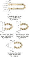
Structures of HIV TAR RNA binding cyclic peptides derived from RNA-recognition motif domains.
FIGURE 7
FIGURE 7
Structures of HIV RRE RNA binding cyclic peptides. n. r. = not reported.
FIGURE 8
FIGURE 8
The steps in the biogenesis of miRNA. Binding to a pre-miRNA can block the pathway and inhibit miRNA downstream effects.
FIGURE 9
FIGURE 9
Structures of (pre-)miRNA and viral RNA binding cyclic peptides. n. r. = not reported. S5 = (s)-pentenylalanine.
FIGURE 10
FIGURE 10
(A) Screening of genetically encoded peptides (i.e., mRNA or phage display). using an immobilized RNA target. (B) Screening of bead-bound synthetic peptides using a fluorescently labelled RNA.
FIGURE 11
FIGURE 11
Structures of RNA-binding cyclic peptides identified via library screening.
FIGURE 12
FIGURE 12
Structures of cyclic peptides natural products that target the ribosome.
FIGURE 13
FIGURE 13
Crystal structures of antibiotic peptides bound to the ribosome. (A) Capreomycin (31) bound to the 16S rRNA (PDB ID—4V7M). (B) Quinupristin (32) bound to the 23S rRNA (PDB ID—4U26). (C) Thiostrepton (33) bound to the 23S rRNA (PDB ID—3CF5).
FIGURE 14
FIGURE 14
Structures of natural product derived cyclic peptides targeting RNA. n. r. = not reported.

Similar articles

Cited by

References

    1. Ahmad A., Ginnebaugh K. R., Sethi S., Chen W., Ali R., Mittal S., et al. (2015). miR-20b Is Up-Regulated in Brain Metastases from Primary Breast Cancers. Oncotarget 6, 12188–12195. 10.18632/oncotarget.3664 - DOI - PMC - PubMed
    1. Allers J., Shamoo Y. (2001). Structure-based Analysis of Protein-RNA Interactions Using the Program ENTANGLE. J. Mol. Biol. 311, 75–86. 10.1006/jmbi.2001.4857 - DOI - PubMed
    1. Athanassiou Z., Dias R. L. A., Moehle K., Dobson N., Varani G., Robinson J. A. (2004). Structural Mimicry of Retroviral Tat Proteins by Constrained β-Hairpin Peptidomimetics: Ligands with High Affinity and Selectivity for Viral TAR RNA Regulatory Elements. J. Am. Chem. Soc. 126, 6906–6913. 10.1021/ja0497680 - DOI - PubMed
    1. Bagley M. C., Dale J. W., Merritt E. A., Xiong X. (2005). Thiopeptide Antibiotics. Chem. Rev. 105, 685–714. 10.1021/cr0300441 - DOI - PubMed
    1. Bai Y., Tambe A., Zhou K., Doudna J. A. (2014). RNA-guided Assembly of Rev-RRE Nuclear export Complexes. eLife 3, 1–17. 10.7554/eLife.03656 - DOI - PMC - PubMed