Review
. 2020 Oct 6;10(60):36751-36777.
doi: 10.1039/d0ra06681h. eCollection 2020 Oct 1.

Advances in chemistry and composition of soft materials for drug releasing contact lenses

Affiliations
Review

Advances in chemistry and composition of soft materials for drug releasing contact lenses

Subir Chatterjee et al. RSC Adv. .

Abstract

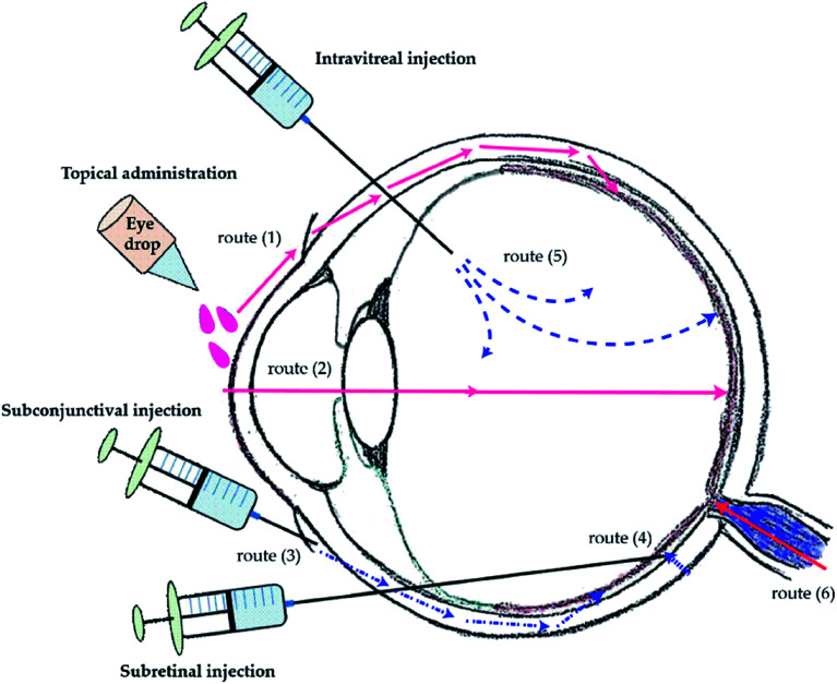
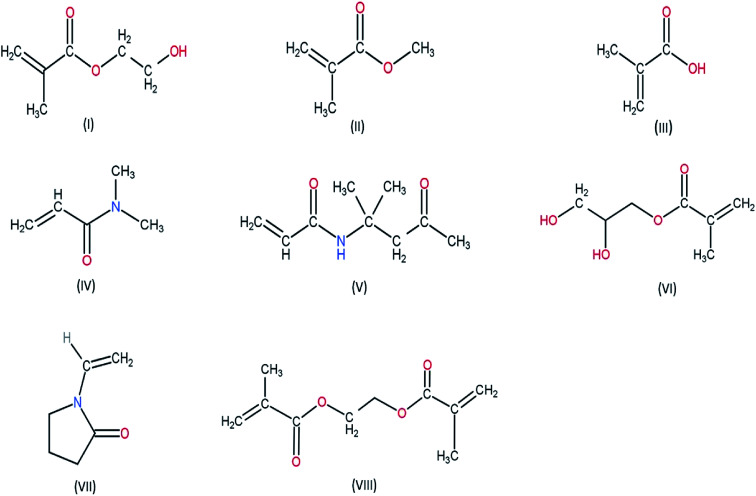
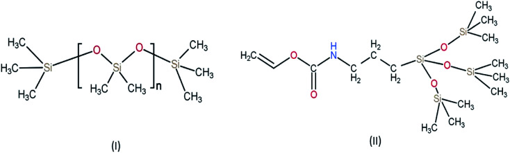
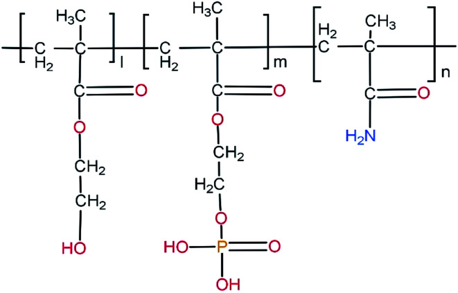
Ocular drug delivery has always been a challenging feat to achieve in the field of medical sciences. One of the existing methods of non-invasive ocular drug delivery is the use of eye drops. However, drugs administered through these formulations have low bioavailability in the ocular system. This limitation can been overcome by using contact lenses as drug delivery vehicles. According to USA FDA definitions they can be categorized into two main categories-hard and soft contact lenses. Based on the material properties, hard contact lenses are mostly produced from polymers of acrylate monomers such as MMA (methyl methacrylate). These have the least water retention capacity, thereby, having minimal ability to diffuse oxygen into the corneal layer and are not ideal for long term use. Soft material contact lenses are flexible and are mainly hydrogel based. They have higher water retention capacities as compared to rigid contact lenses, which gives them the ability to transmit oxygen to the corneal layer. These hydrogel based soft materials are mainly produced from polymers of acrylate monomers such as HEMA (hydroxyethyl methacrylate) and found to be better for drug delivery contact lenses. These polymer-based soft materials have been efficiently modified in terms of their chemistry to achieve diverse physicochemical properties to produce efficient ocular drug delivery systems. However, complications such as drug leaching during storage and distribution, sterilisation, preservation of integrity of the lens and the possibility of surface roughness due to the incorporated drug molecules still need to be optimised. This review highlights the chemistries of various polymeric molecules through which physicochemical properties can be modified to achieve optimum drug loading and sustained release of the drug for application in the ocular system.


Conflict of interest statement

There are no conflicts to declare.

Figures

Fig. 1
Fig. 1. Methods of ocular drug administration and its delivery routes to the posterior segment. Routes of drug transportation to the back of the eye via topical administration (1) and (2), subconjunctival injection (3), subretinal injection (4), and intravitreal injection (5). The drug transportation from the systemic circulation via oral medication (6). Reprinted from ref. 134.
Fig. 2
Fig. 2. Chemical structure of monomers used for various hydrogel based contact lens (I) HEMA (hydroxy ethylmethacrylate) (II) MMA (methyl methacrylate) (III) MAA (methacrylic acid) (IV) DMA (N,N-dimethylacryamide) (V) DAA (diacetone acrylamide) (VI) GMA (glycerol methacrylate) (VII) NVP (N-vinyl pyrrolidone) (VIII) EGDMA (ethylene glycol dimethacrylate).
Fig. 3
Fig. 3. Chemical structure of monomers used for silicone-based hydrogel contact lens (I) PDMS (polydimethylsiloxane) (II) TPVC (tris(trimethylsiloxy)silyl)propyl vinyl carbamate.
Fig. 4
Fig. 4. Effect of loading temperature on the release of dug. Reprinted with permission from ref. 49 copyright (2018), Elsevier.
Fig. 5
Fig. 5. Loading results of betaxolol hydrochloride (BH) (A) and betaxolol base (BB) (B) in NaCl,Na2SO4 and NaSCN solutions with the ionic strength ranging from 0 to 1 mol l−1 and comparison of enhancement factor for diclofenac sodium (DS), betaxolol base and betaxolol hydrochloride in NaCl solutions (C). Each point represents the average of three measurements. (*P < 0.05, **P < 0.01, ***P < 0.001). Reprinted with permission from ref. 51 copyright (2019) Elsevier.
Fig. 6
Fig. 6. Schematic representation of molecular imprinting technique. Reprinted from ref. 63 with permission from the Royal Society of Chemistry.
Fig. 7
Fig. 7. Schematic illustration of lysozyme-activated drug eluting contact lens. (a) Drug loaded ND-nanogels are synthesized by cross-linking PEI-coated NDs and partially N-acetylated chitosan (MW = 57 kDa; degree of N-acetylation = 50%) in the presence of timolol maleate. The ND-nanogels are then embedded in a hydrogel and cast into enzyme-responsive contact lenses. (b) Exposure to lacrimal fluid lysozyme cleaves the N-acetylated chitosan, degrading the ND-nanogels and releasing the entrapped timolol maleate while leaving the lens intact. Reprinted with permission from ref. 72. Copyright 2014, American Chemical Society.
Fig. 8
Fig. 8. Characterization of physical properties of contact lenses. (a) ND-nanogels can be embedded into polyHEMA gels and cast into contact lenses. (b) ND-nanogel embedded lenses maintain optical transparency. (c) Comparison of average visible light transmittance (400–700 nm) and water content of polyHEMA lenses without additives, with 0.1% (w/w) PEI-chitosan, with 0.1% (w/w) pristine ND, with 0.1% (w/w) ND-nanogel or 0.2% (w/w) ND-nanogel. (d) Tensile stress–strain curves comparing polyHEMA lenses without additives, with 0.1% (w/w) PEI-chitosan, with 0.1% (w/w) pristine ND, with 0.1% (w/w) ND-nanogel or 0.2% (w/w) ND-nanogel. (e) Young's modulus of the polyHEMA lenses without additives, with 0.1% (w/w) PEI-chitosan, with 0.1% (w/w) pristine ND, with 0.1% (w/w) ND-nanogel or 0.2% (w/w) ND-nanogel, as determined by the first 5% of the stress–strain curve slope. Reprinted with permission from ref. 72. Copyright 2014, American Chemical Society.
Fig. 9
Fig. 9. Enzyme-triggered drug release. Drug-eluting profiles from drug-soaked (black line), molecularly imprinted (red line) and ND nanogel-embedded contact lenses (blue line) in saline solution at 37 °C as determined by HPLC analysis of TM. Lysozyme (2.7 mg mL−1) in PBS was added after 24 h of incubation (N = 3 for each type of lens). Reprinted with permission from ref. 72. Copyright 2014, American Chemical Society.
Fig. 10
Fig. 10. A schematic representation of the microstructure of the surfactant laden gels. Reprinted with permission from ref. 66. Copyright (2009), Elsevier.
Fig. 11
Fig. 11. Schematic diagram of liposomes immobilized on the surface of contact lens. Reprinted with permission from ref. 82. Copyright (2018), Elsevier.
Fig. 12
Fig. 12. Cyclodextrin-based contact lens used for drug delivery. (a) Copolymerization of acrylic/vinyl CDs derivatives; (b) grafting of CDs to preformed polymer networks; (c) directing cross-linking of CDs. Reprinted with permission from ref. 82. Copyright (2018), Elsevier.
Fig. 13
Fig. 13. Schematic representation of the synthesis of TSC (thiosemicarbazone) loaded beta CD conjugated soft contact lenses by DP (during polymerization) method and PP (post polymerization) method. Reprinted with permission from ref. 95. Copyright (2013), Elsevier.
Fig. 14
Fig. 14. Thiosemicarbazone release kinetics from different hydrogels over two weeks, at 25 °C. (A) pHEMA-co-β-CD produced by the DP method, (B) pHEMA-co-β-CD produced by the PP method and (C) HPβ-CD and HPβ-CD/HPMC SHHs. The artificial lacrimal fluid medium was replaced to maintain sink conditions every (A) 30 h, (B) 4 h and (C) 8 h. Figure insets show the release during the first 24 h. Reprinted with permission from ref. 95. Copyright (2013), Elsevier.
Fig. 15
Fig. 15. Schematic representation of the pH dependent release of drug from pHEMA lens embedded with drug. Reprinted with permission from ref. 102. Copyright (2018), Elsevier.
Fig. 16
Fig. 16. Chemical structure of poly(HEMA-co-MOEP-co-Mam). The presence of phosphate groups in the side chain of the polymer forming hydrogel is seen.

References

    1. Rowe-Rendleman C. L. Durazo S. A. Kompella U. B. Rittenhouse K. D. Di Polo A. Weiner A. L. Grossniklaus H. E. Naash M. I. Lewin A. S. Horsager A. Edelhauser H. F. Drug and gene delivery to the back of the eye: from bench to bedside. Investig. Ophthalmol. Vis. Sci. 2014;55(4):2714–2730. doi: 10.1167/iovs.13-13707. - DOI - PMC - PubMed
    1. Willoughby C. E. Ponzin D. Ferrari S. Lobo A. Landau K. Omidi Y. Anatomy and physiology of the human eye: effects of mucopolysaccharidoses disease on structure and function–a review. Clin. Exp. Ophthalmol. 2010;38:2–11. doi: 10.1111/j.1442-9071.2010.02363.x. - DOI - PubMed
    1. Reimondez-Troitiño S. Csaba N. Alonso M. J. De La Fuente M. Nanotherapies for the treatment of ocular diseases. Eur. J. Pharm. Biopharm. 2015;95:279–293. doi: 10.1016/j.ejpb.2015.02.019. - DOI - PubMed
    1. Quigley H. A. Neuronal death in glaucoma. Prog. Retin. Eye Res. 1999;18(1):39–57. doi: 10.1016/S1350-9462(98)00014-7. - DOI - PubMed
    1. Janoria K. G. Gunda S. Boddu S. H. Mitra A. K. Novel approaches to retinal drug delivery. Expet Opin. Drug Deliv. 2007;4(4):371–388. doi: 10.1517/17425247.4.4.371. - DOI - PubMed