

Effect of small molecular weight soybean protein-derived peptide supplementation on attenuating burn injury-induced inflammation and accelerating wound healing in a rat model
- PMID: 35518054
- PMCID: PMC9059567
- DOI: 10.1039/c8ra09036j
Effect of small molecular weight soybean protein-derived peptide supplementation on attenuating burn injury-induced inflammation and accelerating wound healing in a rat model
Abstract
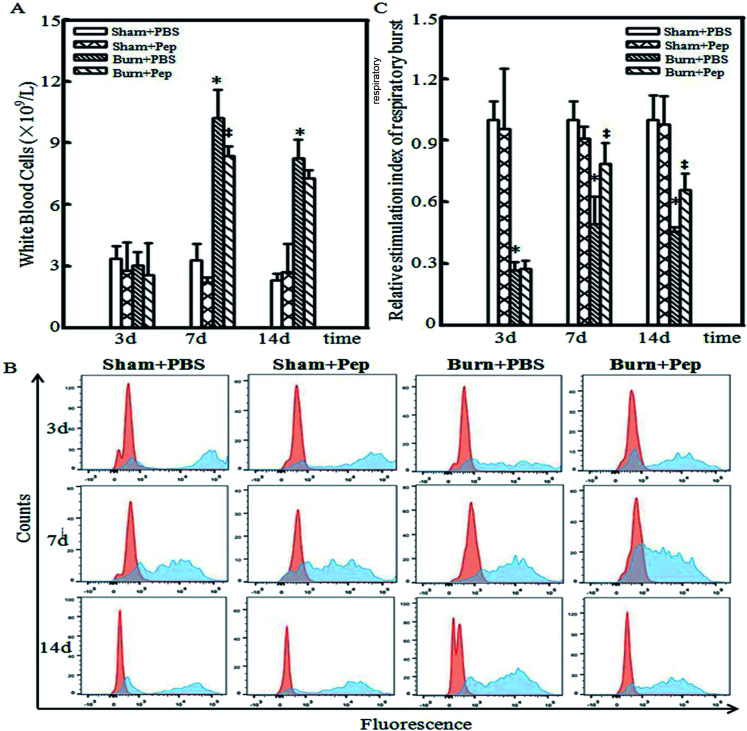
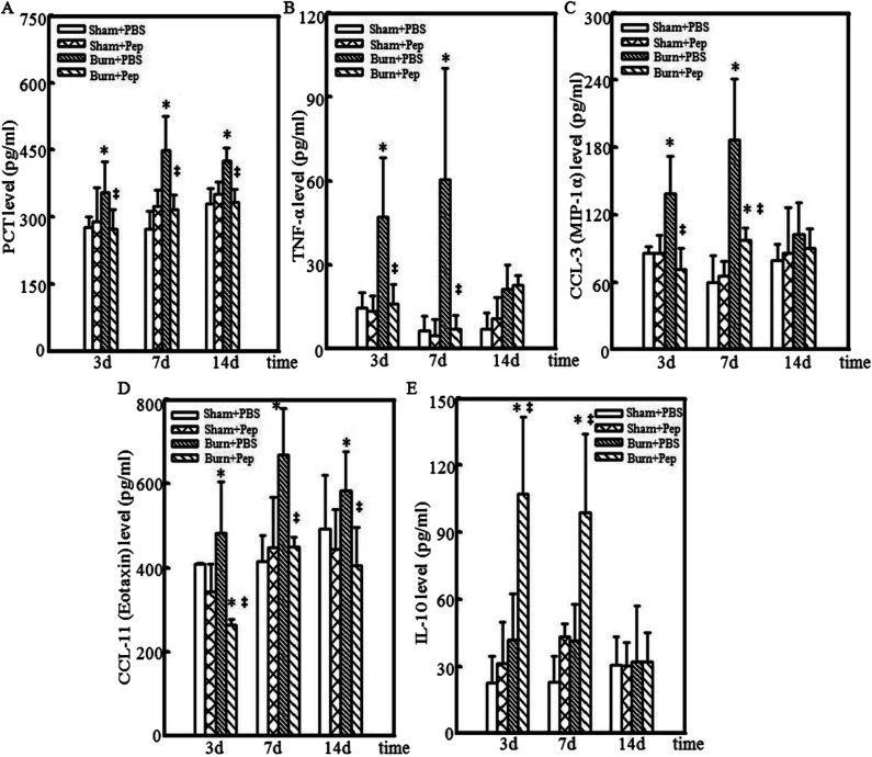
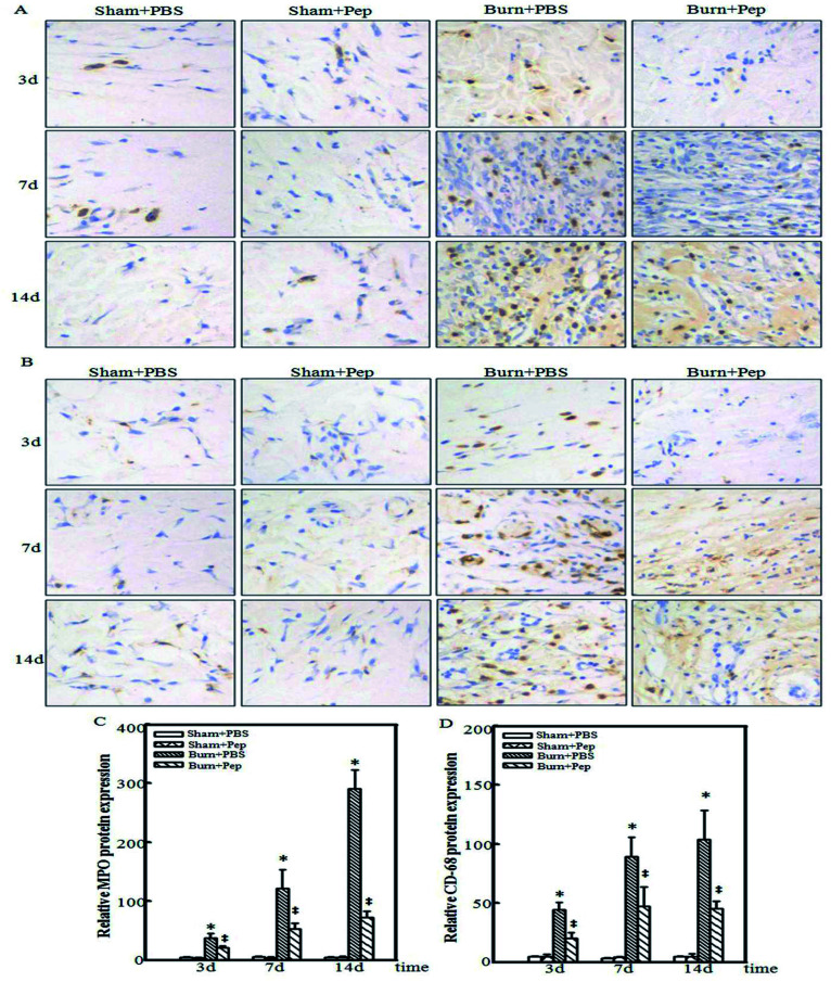
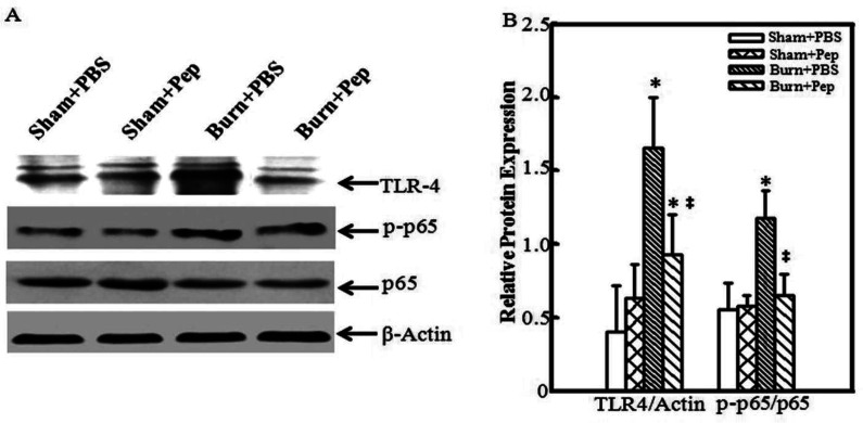
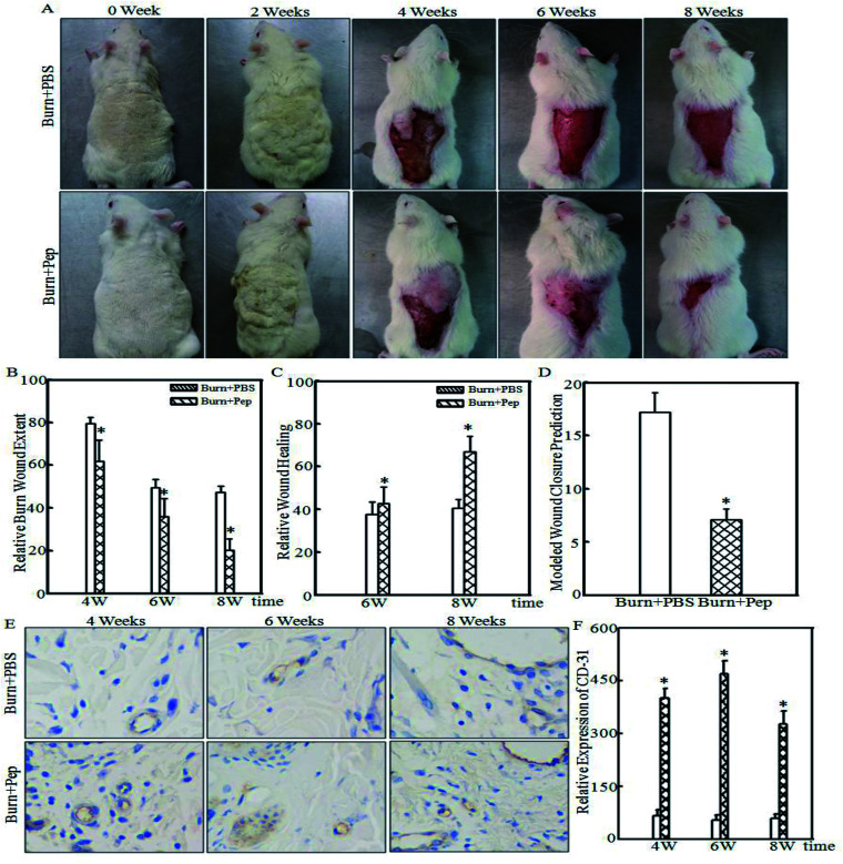
The populations most afflicted by burn injuries have limited abilities to support the significant specialized requirements and costs for acute and long-term burn injury care. This article describes the results of optimizing the use of readily absorbed small molecular weight soybean protein enzymolysis-derived peptide to attenuate rat burn injury-induced inflammation and accelerate wound healing. A major full-thickness 30% total body surface area burn-injury rat model was utilized and the systemic white blood cell (WBC) counts, the relative level of stimulation index of respiratory burst, and the inflammatory markers procalcitonin (PCT), tumor necrosis factor-α (TNF-α), chemokine (C-C motif) ligand 3 (CCL-3), chemokine (C-C motif) ligand 11 (CCL-11) and interleukin-10 (IL-10) were assessed. The burn injury-induced neutrophil and macrophage immune cell infiltration of the cutaneous tissues was detected by immunohistochemical analysis of the protein markers myeloperoxidase (MPO) and cluster of differentiation 68 (CD-68). The local induction of the burn injury-induced toll-like receptor 4/nuclear factor kappa-light-chain-enhancer of activated B (TLR4/NF-κB) signaling pathway in the effected cutaneous tissues was determined by the quantification of the protein expression of TLR4 and phosphorylated NF-κB/p65 using Western blots. In addition, burn wound size and healing rate were assessed biweekly for 8 weeks by imaging and measuring the burn wound surface area, and the angiogenesis protein marker of cluster of differentiation 31 (CD-31) expression in cutaneous tissues was also detected by immunohistochemical analysis. The results showed that nutrient supplementation with optimized readily absorbed small molecular weight soybean protein-derived peptide resulted in a dramatic anti-inflammatory effect as evidenced by the significant increase in the burn injury-induced systemic white blood cell counts and their relative level of stimulation index of respiratory burst, reduction in the burn injury-induced activation of NF-κB transcriptional signaling pathways, significant reduction in the local burn injury-induced cutaneous infiltration of neutrophils and macrophages at all measured time points, reduction in wound size and improved rate of burn injury wound healing with increased CD-31 protein expression. These results indicated that dietary supplementation with small molecular weight soybean-derived peptides could be used as an adjunct therapy in burn injury management to reduce inflammation and improve overall patient outcomes.
This journal is © The Royal Society of Chemistry.
Conflict of interest statement
The authors declare no conflict of interest.
Figures
Similar articles
-
Small Molecular Weight Soybean Protein-Derived Peptides Nutriment Attenuates Rat Burn Injury-Induced Muscle Atrophy by Modulation of Ubiquitin-Proteasome System and Autophagy Signaling Pathway.J Agric Food Chem. 2018 Mar 21;66(11):2724-2734. doi: 10.1021/acs.jafc.7b05387. Epub 2018 Mar 8. J Agric Food Chem. 2018. PMID: 29493231
-
[Effect and mechanism of astaxanthin on acute kidney injury in rats with full-thickness burns].Zhonghua Shao Shang Za Zhi. 2020 Nov 20;36(11):1050-1059. doi: 10.3760/cma.j.cn501120-20200526-00287. Zhonghua Shao Shang Za Zhi. 2020. PMID: 33238688 Chinese.
-
Soybean protein-derived peptides inhibit inflammation in LPS-induced RAW264.7 macrophages via the suppression of TLR4-mediated MAPK-JNK and NF-kappa B activation.J Food Biochem. 2020 Aug;44(8):e13289. doi: 10.1111/jfbc.13289. Epub 2020 Jun 14. J Food Biochem. 2020. PMID: 32537742
-
[Induction mechanism of shock: applying the etiology in judgment of the cause of death in forensic practice].Nihon Hoigaku Zasshi. 2004 Sep;58(2):130-40. Nihon Hoigaku Zasshi. 2004. PMID: 15526767 Review. Japanese.
-
Burn injury induces elevated inflammatory traffic: the role of NF-κB.Inflamm Res. 2021 Jan;70(1):51-65. doi: 10.1007/s00011-020-01426-x. Epub 2020 Nov 27. Inflamm Res. 2021. PMID: 33245371 Review.
Cited by
-
Soybean protein-derived peptide nutriment increases negative nitrogen balance in burn injury-induced inflammatory stress response in aged rats through the modulation of white blood cells and immune factors.Food Nutr Res. 2020 Jun 29;64. doi: 10.29219/fnr.v64.3677. eCollection 2020. Food Nutr Res. 2020. PMID: 32694965 Free PMC article.
-
The Beneficial Effects of Soybean Proteins and Peptides on Chronic Diseases.Nutrients. 2023 Apr 7;15(8):1811. doi: 10.3390/nu15081811. Nutrients. 2023. PMID: 37111030 Free PMC article. Review.
-
Investigating Differential Expressed Genes of Limosilactobacillus reuteri LR08 Regulated by Soybean Protein and Peptides.Foods. 2022 Apr 26;11(9):1251. doi: 10.3390/foods11091251. Foods. 2022. PMID: 35563974 Free PMC article.
-
Effect of blended protein nutritional support on reducing burn-induced inflammation and organ injury.Nutr Res Pract. 2022 Oct;16(5):589-603. doi: 10.4162/nrp.2022.16.5.589. Epub 2022 Apr 12. Nutr Res Pract. 2022. PMID: 36238375 Free PMC article.
-
Statins block mammalian target of rapamycin pathway: a possible novel therapeutic strategy for inflammatory, malignant and neurodegenerative diseases.Inflammopharmacology. 2023 Feb;31(1):57-75. doi: 10.1007/s10787-022-01077-w. Epub 2022 Dec 27. Inflammopharmacology. 2023. PMID: 36574095 Free PMC article. Review.
References
-
- World Health Organization, Global health estimates 2015: Disease burden by cause, age, sex, by country and by region, 2000–2015, Geneva, 2016
-
- Pruitt Jr B. A., Wolf S. E. and Mason Jr A. D., in Total Burn Care, W.B. Saunders, London, 4th edn, 2012, pp. 15–45.e14
-
- Tintinalli J., Emergency Medicine (Tintinalli), New York, 2010, pp. 1374–1386
LinkOut - more resources
Full Text Sources
Other Literature Sources
Research Materials
Miscellaneous