. 2019 Jan 29;9(7):3740-3748.
doi: 10.1039/c8ra09171d. eCollection 2019 Jan 25.

A Col I and BCP ceramic bi-layer scaffold implant promotes regeneration in osteochondral defects

Affiliations

A Col I and BCP ceramic bi-layer scaffold implant promotes regeneration in osteochondral defects

Hanxu Cai et al. RSC Adv. .

Abstract

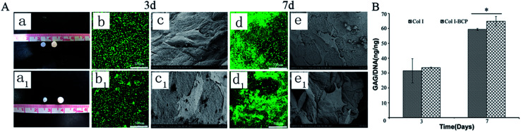
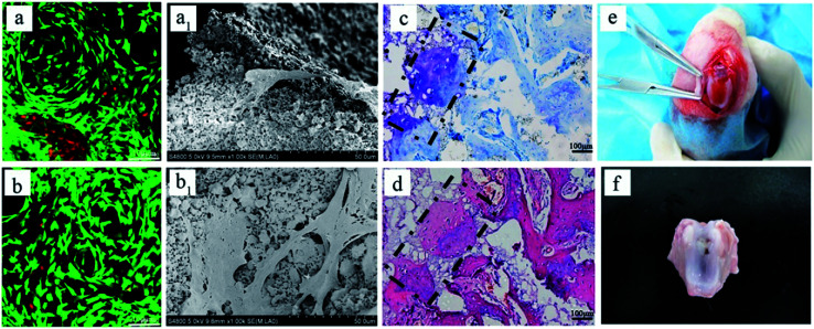
Osteochondral defects occur in the superficial cartilage region, intermediate calcified cartilage, and subchondral bone. Due to the limited regenerative capacity and complex zonal structure, it is critically difficult to develop strategies for osteochondral defect repair that could meet clinical requirements. In this study, type I collagen (Col I) and BCP ceramics were used to fabricate a new bi-layer scaffold for regeneration in osteochondral defects. The in vitro studies showed that the bi-layer scaffold provided special functions for cell migration, proliferation and secretion due to the layered scaffold structure. The in vivo results demonstrated that the bi-layered scaffold could effectively promote the regeneration of both the cartilage and the subchondral bone, and the newly formed cartilage layer, with a similar structure and thickness to the natural cartilaginous layer, could seamlessly integrate with the surrounding natural cartilage and regenerate an interface layer to mimic the native osteochondral structure.


Conflict of interest statement

There are no conflicts to declare.

Figures

Fig. 1
Fig. 1. (A) Structure of the bi-layer scaffolds observed by SEM. (B) Morphological changes of Col I (a1) and bi-layer (b2) scaffolds after immersion in PBS for 7 days. (C) The equilibrium swelling ratio change after immersion in PBS at pH 7.4 and 37 °C.
Fig. 2
Fig. 2. The Col I hydrogel and bi-layer scaffolds with cultured chondrocytes (5 × 10⁶ cells per mL) in vitro. (A) Macroscopic morphology changes of Col I hydrogel (left) and bi-layer scaffold (right) after 3 (a) and 7 (a1) days of culture. The CLSM micrographs of chondrocytes encapsulated in Col I hydrogel after culture for 3 (b) and 7 (d) days, and the in bi-layer scaffold after culture for 3 days (b1) and 7 days (d1). The SEM micrographs of chondrocytes encapsulated in Col I hydrogel after culture for 3 days (c) and 7 days (e), and in bi-layer scaffold after culture for 3 days (c1) and 7 days (e1). (B) The quantification of GAG content secreted by chondrocytes after 3 and 7 days culture. *p < 0.05.
Fig. 3
Fig. 3. Scaffolds were implanted into defects for in vivo treatment of osteochondral defects. (A) Osteochondral defects were created on the patellar trochlear groove and then filled with different scaffolds or no treatment as control (a–d). Gross and section morphology after 2 weeks (e–h and e1–h1) and 4 weeks (i–l and i1–l1). (B) Macroscopic evaluation according to ICRS macroscopic scores. *p < 0.05, **p < 0.01, ***p < 0.001.
Fig. 4
Fig. 4. (A) HE staining to analyze subchondral bone repair. (B) The density of regenerated bone at 2 and 4 weeks was calculated by Image-Pro Plus software. — new bone, —osteoid, —blood vessel. *p < 0.05, **p < 0.01, ***p < 0.001.
Fig. 5
Fig. 5. (A) HE, TB and Saf.O staining images show the regeneration status of tissues after 2 weeks. (B) Cartilage thickness was calculated by Image-Pro Plus software. The dashed line and arrows indicate the collapse of cartilage due to absence of subchondral bone support. — the surface of cartilage, *p < 0.05, **p < 0.01, ***p < 0.001.
Fig. 6
Fig. 6. (A) HE, TB and Saf.O staining images show the regeneration status of tissue after 4 weeks. (B) Cartilage thickness was calculated using Image-Pro Plus software. The green dashed boxes indicate the connection between the cartilage and the subchondral bone. The black dashed boxes indicate the connection between the newly formed cartilage tissue and surrounding healthy cartilage. — the surface of cartilage, *p < 0.05, **p < 0.01, ***p < 0.001.
Fig. 7
Fig. 7. The CLSM micrographs of chondrocytes encapsulated in bi-layer scaffold after culture for (a) 3 and (b) 7 days. The SEM micrographs of chondrocytes encapsulated in bi-layer scaffold after culture for (a1) 3 and (b1) 7 days. (c) TB and (d) HE staining images show the regeneration status of cartilage–bone interface after 4 weeks. During operation, (e) bleeding affected type I collagen gelation and (f) blood clot affected the repair effect of the cartilage layer.

Similar articles

Cited by

References

    1. Nukavarapu S. P. Dorcemus D. L. Biotechnol. Adv. 2013;31:706. doi: 10.1016/j.biotechadv.2012.11.004. - DOI - PubMed
    1. Sartori M. Pagani S. Ferrari A. Costa V. Carina V. Figallo E. Maltarello M. C. Martini L. Fini M. Giavaresi G. Mater. Sci. Eng., C. 2017;70:101. doi: 10.1016/j.msec.2016.08.027. - DOI - PubMed
    1. Chen P. Xia C. Mo J. Mei S. Lin X. Fan S. Mater. Sci. Eng., C. 2018;91:190. doi: 10.1016/j.msec.2018.05.034. - DOI - PubMed
    1. Hunter D. J. Osteoarthritis Cartilage. 2009;17:1402. doi: 10.1016/j.joca.2009.04.014. - DOI - PubMed
    1. Lu S. Lam J. Trachtenberg J. E. Lee E. J. Seyednejad H. van den Beucken J. Tabata Y. Wong M. E. Jansen J. A. Mikos A. G. Kasper F. K. Biomaterials. 2014;35:8829. doi: 10.1016/j.biomaterials.2014.07.006. - DOI - PMC - PubMed