. 2020 Aug 26;10(53):31748-31757.
doi: 10.1039/d0ra05948j.

Boron Schiff bases derived from α-amino acids as nucleoli/cytoplasm cell-staining fluorescent probes in vitro

Affiliations

Boron Schiff bases derived from α-amino acids as nucleoli/cytoplasm cell-staining fluorescent probes in vitro

Jesús A Lara-Cerón et al. RSC Adv. .

Abstract

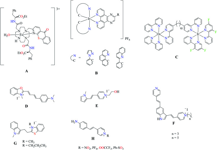
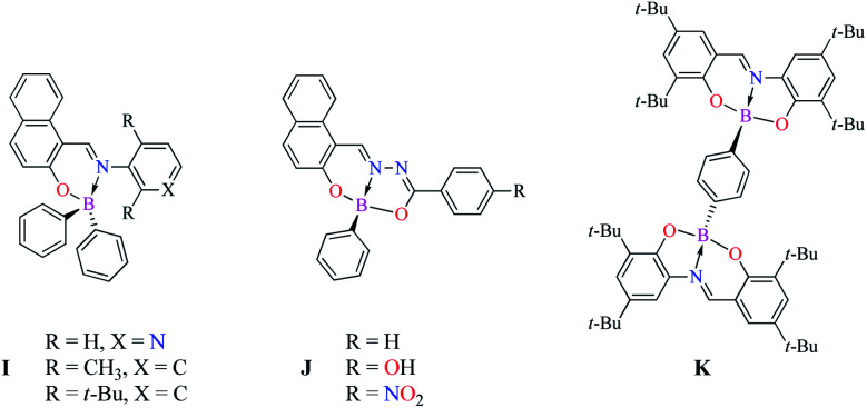
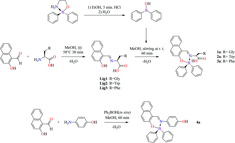
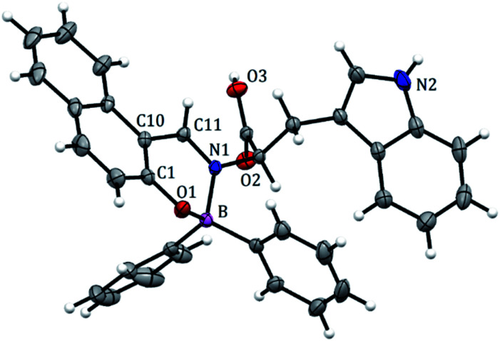
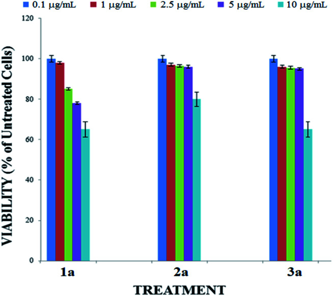
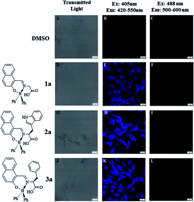
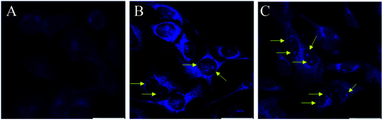
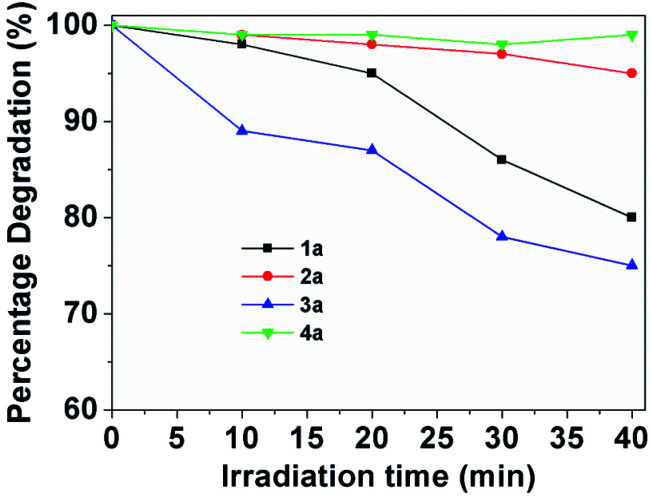
The size, shape, and number of nucleoli in a cell's nucleus might help to distinguish a malignant from a benign tumor. Cellular biology and histopathology often require better visualization to understand nucleoli-related processes, thus organelle-specific fluorescent markers are needed. Here, we report the design, synthesis, and fully chemo-photophysical characterization of fluorescent boron Schiff bases (BOSCHIBAs), derived from α-amino acids (i.e., phenylalanine, tyrosine and tryptophan), with nucleoli- and cytoplasm-specific staining in cells. It is the first time that Boron Schiff bases derived from α-amino acids act as notorious dual (nucleoli and cytoplasm) cell-staining fluorescent probes. The boron derivatives not only showed good photostability and acceptable quantum yields (∼5%) in solution, but also exhibited low cytotoxicity (>90% cell viability at 0.1 and 1 μg mL-1), which make them good candidates to be used in medical diagnosis.


Conflict of interest statement

There are no conflicts to declare.

Figures

Scheme 1
Scheme 1. Fluorescent dyes for in vitro nucleoli staining.
Scheme 2
Scheme 2. Mono and binuclear Boron Schiff bases.
Scheme 3
Scheme 3. Synthesis of Schiff bases by ultrasound Lig1–3 and BOSCHIBAs 1a–4a.
Fig. 1
Fig. 1. Crystal structure of BOSCHIBA 3a. Anisotropic displacement parameters are depicted at the 30% probability level. Hydrogen atoms were omitted for clarity. Distances: B(1)–N(1) 1.631, B(1)–O(1) 1.495, B(1)–C(17) 1.614, B(1)–C(23) 1.618, C(11)–N(1) 1.299 Å. Bond angles: C(11)–B(1)–N(1) 118.64, O(1)–B(1)–N(1) 104.22, O(1)–B(1)–C(17) 110.89, N(1)–B(1)–C(23) 105.98, C(2)–C(11)–N(1) 122.85, C(11)–N(1)–C(12) 119.38°.
Fig. 2
Fig. 2. Intermolecular hydrogen interactions in BOSCHIBA 3a.
Fig. 3
Fig. 3. Boron out of the plane in half chair conformation.
Fig. 4
Fig. 4. Absorption spectra of BOSCHIBAS 1a–4a (short dash dot) and (inset) amino acid Schiff base Lig1–3 (solid lines) in methanol.
Fig. 5
Fig. 5. Emission spectra of BOSCHIBAs 1a–4a and (inset) α-amino acid Schiff bases Lig1–3 in MeOH.
Fig. 6
Fig. 6. Cytotoxicity effect of BOSCHIBAs 1a–3a. B16F10 melanoma cells were treated with 10 mg mL−1 (soft blue bars), 5 mg mL−1 (purple bars), 2.5 mg mL−1 (green bars), 1 mg mL−1 (red bars) or 0.1 mg mL−1 (sharp blue bars) for 24 hours.
Fig. 7
Fig. 7. Confocal fluorescence images of B16F10 melanoma cells stained with BOSCHIBAs 1a–3a (10 μg mL−1). (A–C) Untreated cells; (D–F) compound 1a; (G–I) compound 2a; (J–L) compound 3a (scale bar shown represents 20 mm).
Fig. 8
Fig. 8. Confocal fluorescence images of B16F10 melanoma cells stained with 10 μg mL−1 of BOSCHIBAs 1a (A), 2a, (B), and 3a (C).
Fig. 9
Fig. 9. Photostability of 1a–4a (1 mg/50 mL) in methanol at the absorption maximum wavelength with the irradiation time.

Similar articles

Cited by

References

    1. Hu F. Liu B. Org. Biomol. Chem. 2016;14:9931–9944. doi: 10.1039/C6OB01414C. - DOI - PubMed
    1. Zhu H. Fan J. Du J. Peng X. Acc. Chem. Res. 2016;49(10):2115–2126. doi: 10.1021/acs.accounts.6b00292. - DOI - PubMed
    1. Zheng M. Librizzi D. Kiliç A. Liu Y. Renz H. Merkel O. M. Kissel T. Biomaterials. 2012;33:6551–6558. doi: 10.1016/j.biomaterials.2012.05.055. - DOI - PubMed
    1. Yang Z. Cao J. He Y. Yang J. H. Kim T. Peng X. Kim J. S. Chem. Soc. Rev. 2014;43:4563–4601. doi: 10.1039/C4CS00051J. - DOI - PubMed
    1. Liu H. W. Chen L. Xu C. Li Z. Zhang H. Zhang X. B. Tan W. Chem. Soc. Rev. 2018;47:7140–7180. doi: 10.1039/C7CS00862G. - DOI - PubMed