

Photocatalytic and bactericidal properties and molecular docking analysis of TiO2 nanoparticles conjugated with Zr for environmental remediation
- PMID: 35518250
- PMCID: PMC9056309
- DOI: 10.1039/d0ra05862a
Photocatalytic and bactericidal properties and molecular docking analysis of TiO2 nanoparticles conjugated with Zr for environmental remediation
Abstract
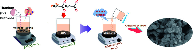
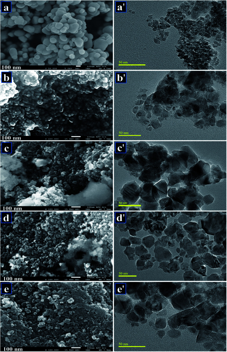
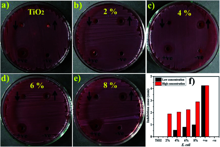
Despite implementing several methodologies including a combination of physical, chemical and biological techniques, aquatic and microbial pollution remains a challenge to this day. Recently, nanomaterials have attracted considerable attention due to their extraordinary prospective for utilization toward environmental remediation. Among several probable candidates, TiO2 stands out due to its potential for use in multifaceted applications. One way to improve the catalytic and antimicrobial potential of TiO2 is to dope it with certain elements. In this study, Zr-doped TiO2 was synthesized through a sol-gel chemical method using various dopant concentrations (2, 4, 6, and 8 wt%). Surface morphological, microstructural and elemental analysis was carried out using FESEM and HR-TEM along with EDS to confirm the formation of Zr-TiO2. XRD spectra showed a linear shift of the (101) anatase peak to lower diffraction angles (from 25.4° to 25.08°) with increasing Zr4+ concentration. Functional groups were examined via FTIR, an ample absorption band appearing between 400 and 700 cm-1 in the acquired spectrum was attributed to the vibration modes of the Ti-O-Ti linkage present within TiO2 nanoparticles, which denotes the formation of TiO2. Experimental results indicated that with increasing dopant concentrations, photocatalytic potential was enhanced significantly. In this respect, TiO2 doped with 8 wt% Zr (sample 0.08 : 1) exhibited outstanding performance by realizing 98% elimination of synthetic MB in 100 minutes. This is thought to be due to a decreased rate of electron-hole pair recombination that transpires upon doping. Therefore, it is proposed that Zr-doped TiO2 can be used as an effective photocatalyst material for various environmental and wastewater treatment applications. The good docking scores and binding confirmation of Zr-doped TiO2 suggested doped nanoparticles as a potential inhibitor against selected targets of both E. coli and S. aureus. Hence, enzyme inhibition studies of Zr-doped TiO2 NPs are suggested for further confirmation of these in silico predictions.
This journal is © The Royal Society of Chemistry.
Conflict of interest statement
There are no conflicts to declare.
Figures


Similar articles
-
Dye degradation performance, bactericidal behavior and molecular docking analysis of Cu-doped TiO2 nanoparticles.RSC Adv. 2020 Jun 25;10(41):24215-24233. doi: 10.1039/d0ra04851h. eCollection 2020 Jun 24. RSC Adv. 2020. PMID: 35516171 Free PMC article.
-
TiO2 Co-doped with Zr and Ag shows highly efficient visible light photocatalytic behavior suitable for treatment of polluted water.RSC Adv. 2020 Nov 19;10(69):42235-42248. doi: 10.1039/d0ra08718a. eCollection 2020 Nov 17. RSC Adv. 2020. PMID: 35516777 Free PMC article.
-
Physical properties and photocatalytic activity of Cr-doped TiO2 nanoparticles.J Microsc. 2023 Sep;291(3):210-228. doi: 10.1111/jmi.13211. Epub 2023 Jul 6. J Microsc. 2023. PMID: 37357432
-
Promising performance of chemically exfoliated Zr-doped MoS2 nanosheets for catalytic and antibacterial applications.RSC Adv. 2020 May 29;10(35):20559-20571. doi: 10.1039/d0ra02458a. eCollection 2020 May 27. RSC Adv. 2020. PMID: 35517731 Free PMC article.
-
A mini-review on rare earth metal-doped TiO2 for photocatalytic remediation of wastewater.Environ Sci Pollut Res Int. 2016 Aug;23(16):15941-51. doi: 10.1007/s11356-016-6984-7. Epub 2016 Jun 22. Environ Sci Pollut Res Int. 2016. PMID: 27335012 Review.
Cited by
-
One-step high-yield preparation of nitrogen- and sulfur-codoped carbon dots with applications in chromium(vi) and ascorbic acid detection.RSC Adv. 2022 Jul 13;12(30):19686-19694. doi: 10.1039/d2ra01758j. eCollection 2022 Jun 29. RSC Adv. 2022. PMID: 35919374 Free PMC article.
-
Impact of Bi Doping into Boron Nitride Nanosheets on Electronic and Optical Properties Using Theoretical Calculations and Experiments.Nanoscale Res Lett. 2021 May 12;16(1):82. doi: 10.1186/s11671-021-03542-x. Nanoscale Res Lett. 2021. PMID: 33978872 Free PMC article.
-
A review on bismuth oxyhalide based materials for photocatalysis.Nanoscale Adv. 2021 May 3;3(12):3353-3372. doi: 10.1039/d1na00223f. eCollection 2021 Jun 15. Nanoscale Adv. 2021. PMID: 36133717 Free PMC article. Review.
-
Development of Multi-concentration Cu:Ag Bimetallic Nanoparticles as a Promising Bactericidal for Antibiotic-Resistant Bacteria as Evaluated with Molecular Docking Study.Nanoscale Res Lett. 2021 May 22;16(1):91. doi: 10.1186/s11671-021-03547-6. Nanoscale Res Lett. 2021. PMID: 34021844 Free PMC article.
-
Using biologically synthesized TiO2 nanoparticles as potential remedy against multiple drug resistant Staphylococcus aureus of bovine mastitis.Sci Rep. 2023 Nov 1;13(1):18785. doi: 10.1038/s41598-023-45762-4. Sci Rep. 2023. PMID: 37914792 Free PMC article.
References
-
- Wypych A. Bobowska I. Tracz M. Opasinska A. Kadlubowski S. Krzywania-Kaliszewska A. Grobelny J. Wojciechowski P. J. Nanomater. 2014;2014:124814. doi: 10.1155/2014/124814. - DOI
-
- Abazari R. Mahjoub A. R. Sanati S. RSC Adv. 2014;4:56406–56414. doi: 10.1039/C4RA10018B. - DOI
-
- Cui Y. Zhang L. Lv K. Zhou G. Wang Z.-S. J. Mater. Chem. A. 2015;3:4477–4483. doi: 10.1039/C4TA06679K. - DOI
LinkOut - more resources
Full Text Sources
Miscellaneous