

High-throughput in-focus differential interference contrast imaging of three-dimensional orientations of single gold nanorods coated with a mesoporous silica shell
- PMID: 35518257
- PMCID: PMC9056269
- DOI: 10.1039/d0ra04704j
High-throughput in-focus differential interference contrast imaging of three-dimensional orientations of single gold nanorods coated with a mesoporous silica shell
Abstract
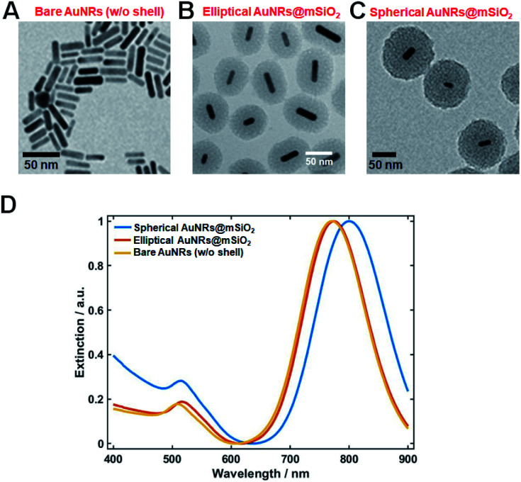
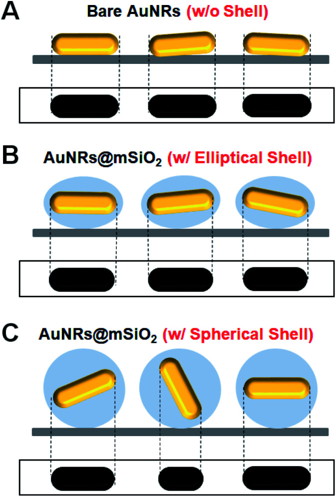
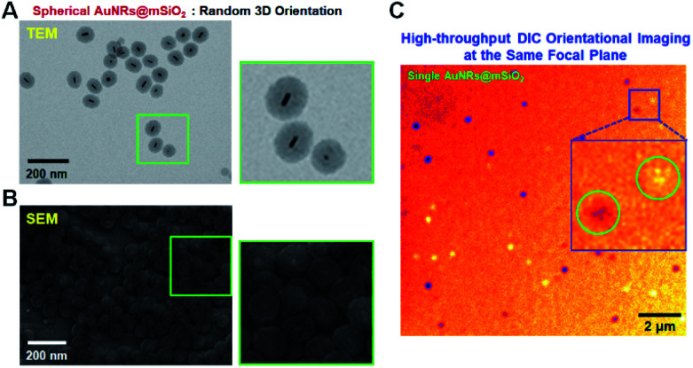
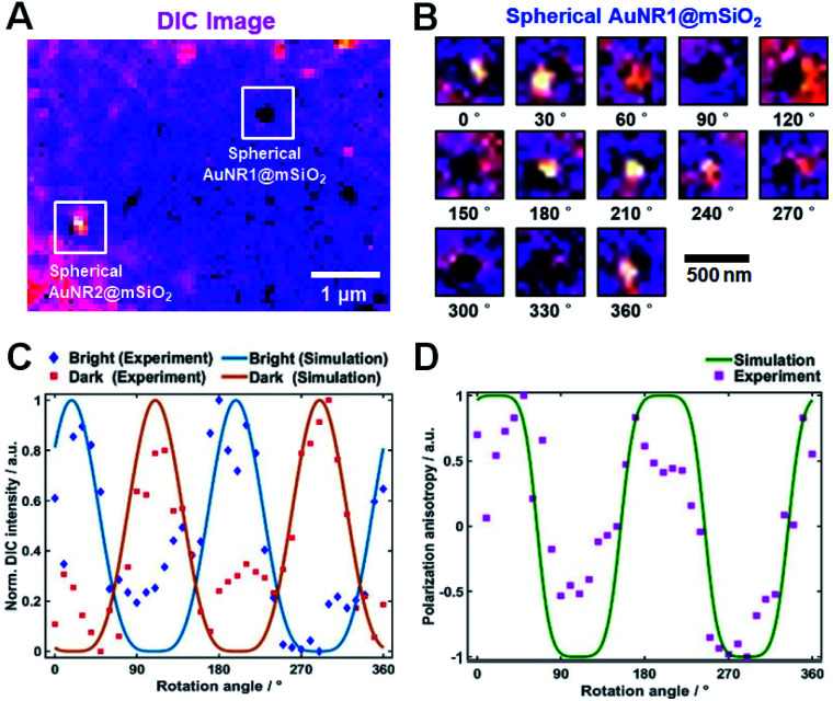
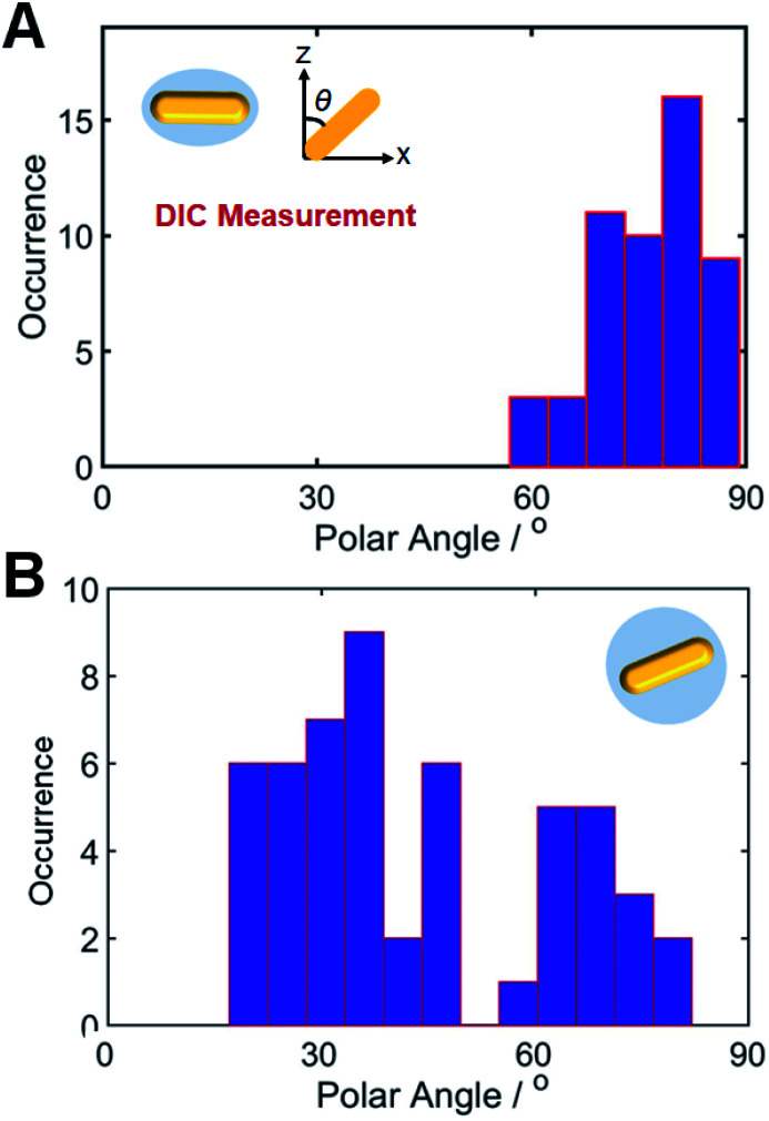
Plasmonic gold nanorods (AuNRs) have been widely applied as optical orientation probes in many biophysical studies. However, characterizing the various three-dimensional (3D) orientations of AuNRs in the same focal plane of the objective lens is a challenging task. To overcome this challenge, we fabricated single AuNRs (10 nm × 30 nm) coated with either an elliptical or spherical mesoporous silica shell (AuNRs@mSiO2). Unlike bare AuNRs and elliptical AuNRs@mSiO2, spherical AuNRs@mSiO2 contained randomly oriented AuNR cores in 3D space, which could be observed on the same focal plane within a single frame by differential interference contrast (DIC) microscopy. The spherical AuNRs@mSiO2 thus achieved high-throughput detection. The proposed approach can overcome the limitations of the current gel-matrix method, which requires vertical scanning of the embedded AuNRs to capture different focal planes.
This journal is © The Royal Society of Chemistry.
Conflict of interest statement
There are no conflicts to declare.
Figures
Similar articles
-
Mesoporous silica shell-coated single gold nanorods as multifunctional orientation probes in dynamic biological environments.RSC Adv. 2021 Dec 1;11(61):38632-38637. doi: 10.1039/d1ra06572f. eCollection 2021 Nov 29. RSC Adv. 2021. PMID: 35493222 Free PMC article.
-
Tuning plasmonic properties by promoting the inward Hg diffusion via oxygen plasma treatment in gold nanorods coated with a mesoporous silica shell.Analyst. 2022 Aug 8;147(16):3623-3627. doi: 10.1039/d2an01007k. Analyst. 2022. PMID: 35861607
-
Single-Particle Study on Hg Amalgamation Mechanism and Slow Inward Diffusion in Mesoporous Silica-Coated Gold Nanorods without Structural Deformation.J Phys Chem Lett. 2022 Mar 24;13(11):2607-2613. doi: 10.1021/acs.jpclett.2c00189. Epub 2022 Mar 16. J Phys Chem Lett. 2022. PMID: 35293762
-
Influence of oxygen plasma treatment on structural and spectral changes in silica-coated gold nanorods studied using total internal reflection microscopy and spectroscopy.Analyst. 2021 Jun 28;146(13):4125-4129. doi: 10.1039/d1an00592h. Analyst. 2021. PMID: 34076657
-
Graphene Oxide-Coated Gold Nanorods: Synthesis and Applications.Nanomaterials (Basel). 2020 Oct 28;10(11):2149. doi: 10.3390/nano10112149. Nanomaterials (Basel). 2020. PMID: 33126610 Free PMC article. Review.
Cited by
-
Direct Observation of In-Focus Plasmonic Cargos via Breaking Angular Degeneracy in Differential Interference Contrast Microscopy.JACS Au. 2023 Dec 2;3(12):3436-3445. doi: 10.1021/jacsau.3c00594. eCollection 2023 Dec 25. JACS Au. 2023. PMID: 38155657 Free PMC article.
References
LinkOut - more resources
Full Text Sources