. 2022 Apr 18:11:e00305.
doi: 10.1016/j.ohx.2022.e00305. eCollection 2022 Apr.

Open source fraction collector/MALDI spotter for proteomics

Affiliations

Open source fraction collector/MALDI spotter for proteomics

Scott B Ficarro et al. HardwareX. .

Abstract

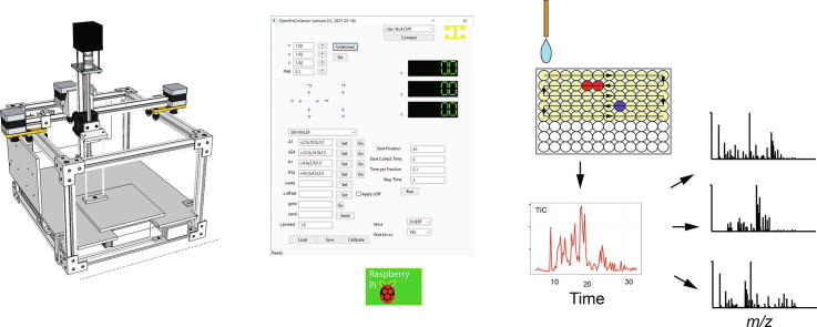
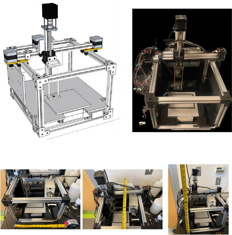
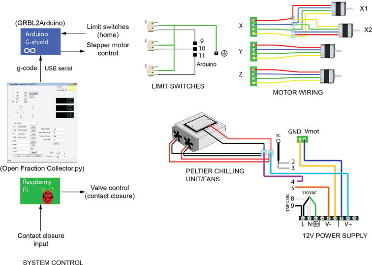
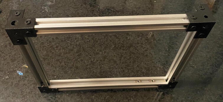
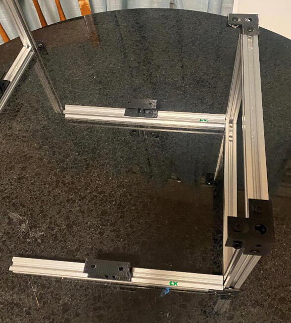
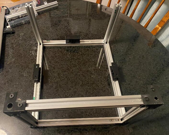
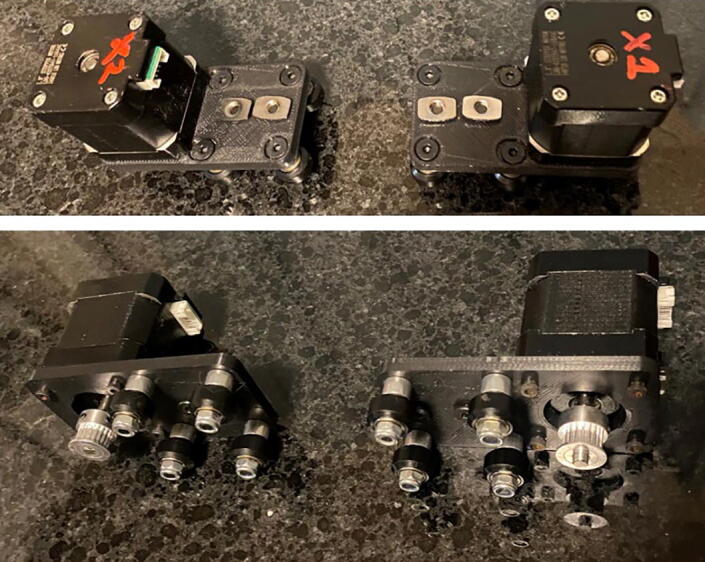
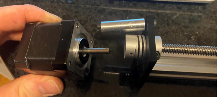
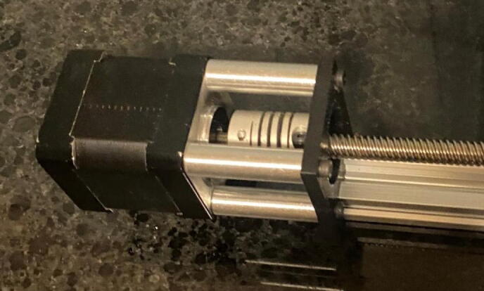
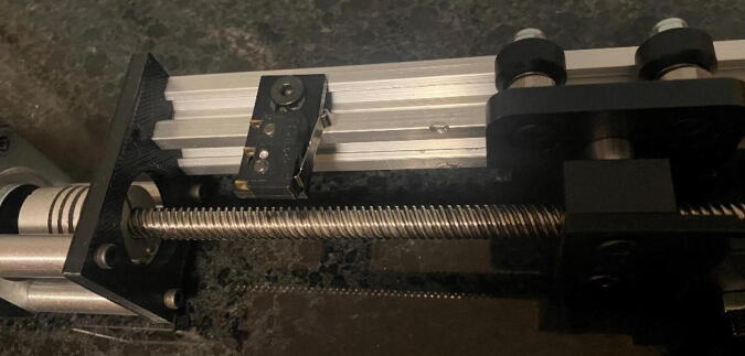
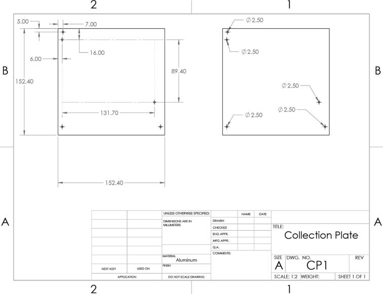
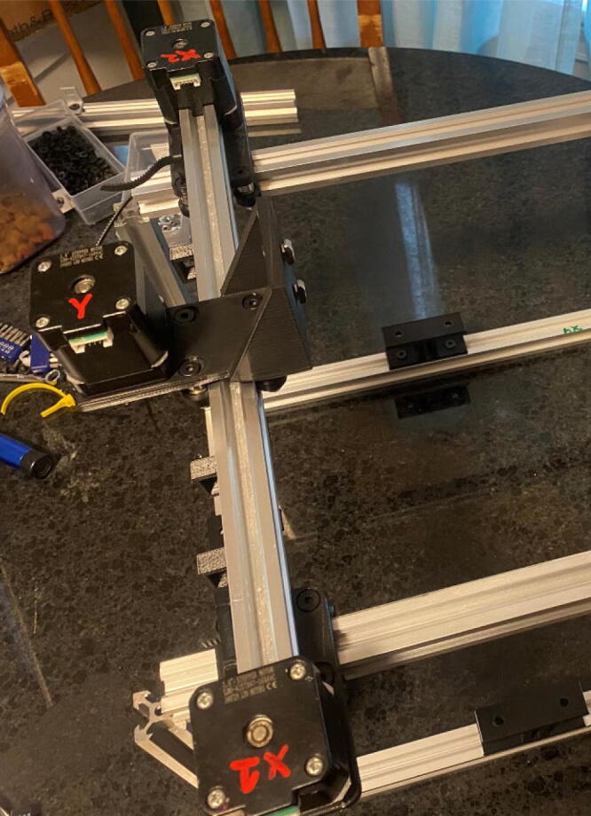
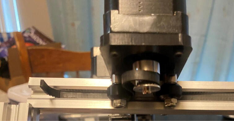
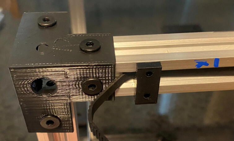
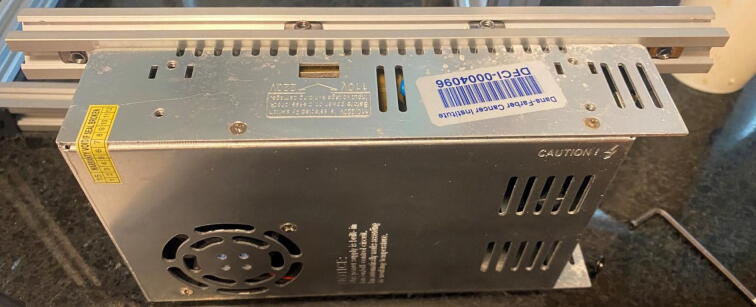
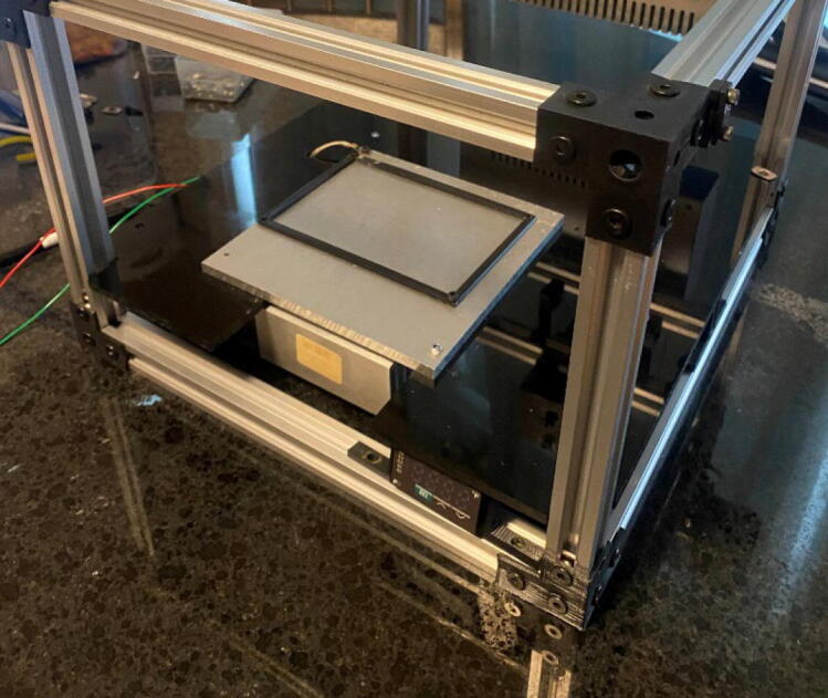
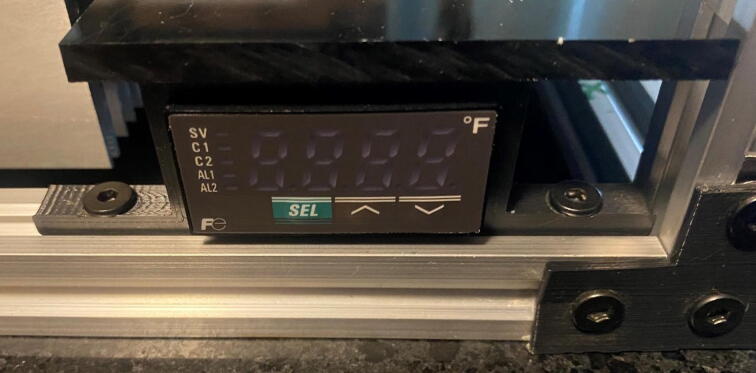
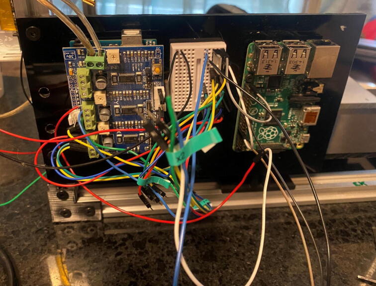
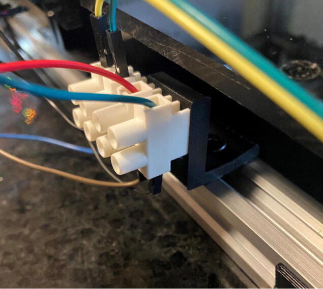
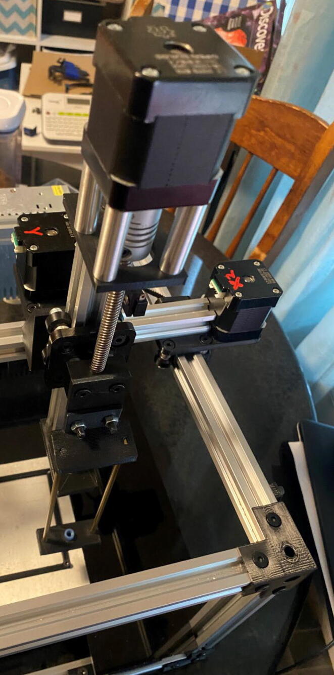
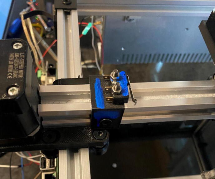
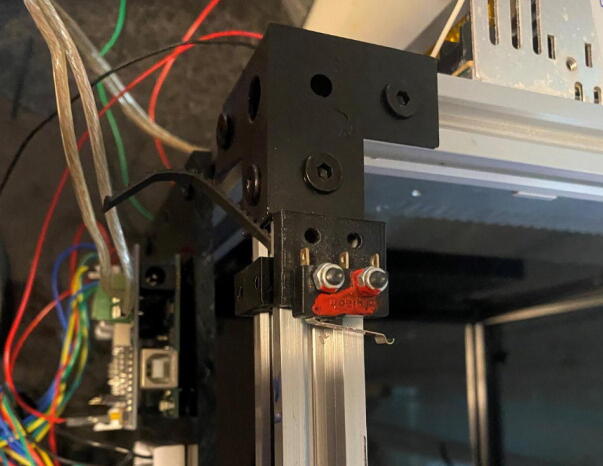
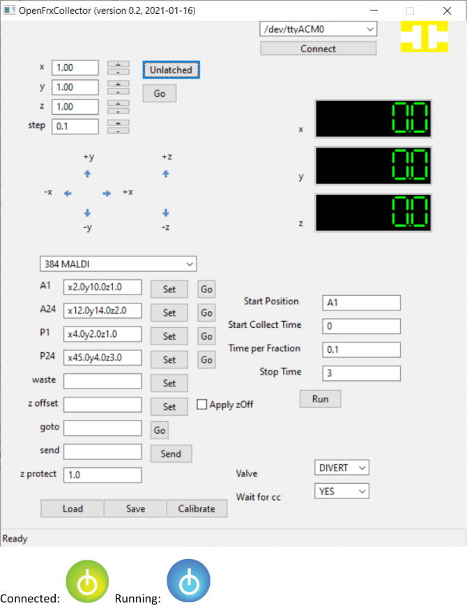
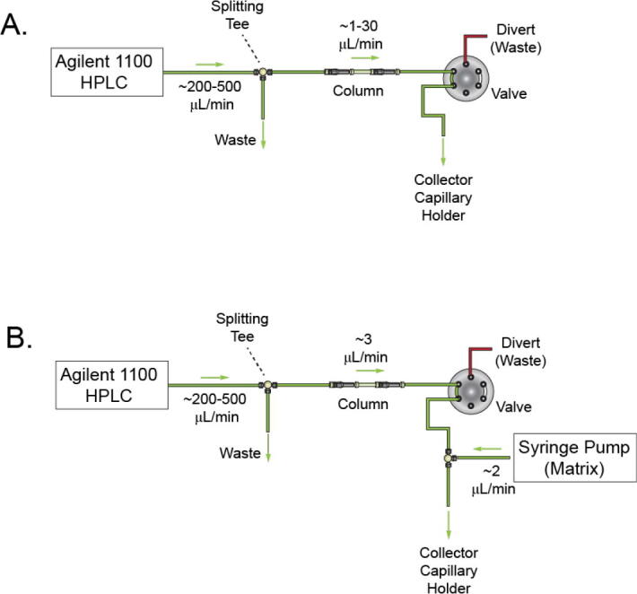
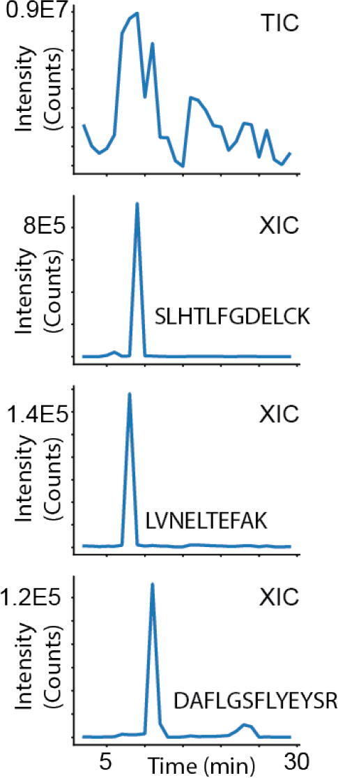
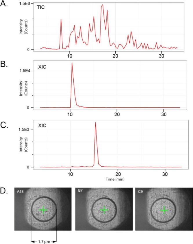
We describe a complete open-source hardware/software solution for high performance thermostatted peptide fraction collection to support mass spectrometry experiments with complex proteomes. The instrument is easy to assemble using parts readily available through retail channels at a fraction of the cost compared to typical commercial systems. Control software is written in Python allowing for rapid customization. We demonstrate several useful applications, including the automated deposition of LC separated peptides for matrix-assisted laser desorption ionization mass spectrometry (MALDI-MS) as well as collection and concatenation of peptide fractions from nanoflow HPLC separations.

Keywords: Fraction collection; HPLC, high pressure liquid chromatography; LC, liquid chromatography; LC-MALDI; LC-MS, Liquid Chromatography/Mass spectrometry; MALDI-MS, matrix-assisted laser desorption ionization mass spectrometry; MS, mass spectrometer/mass spectrometry; OEM, Original Equipment Manufacturer; Open source; Proteomics; Python.


Conflict of interest statement

The authors declare that they have no known competing financial interests or personal relationships that could have appeared to influence the work reported in this paper.

Figures

None
Graphical abstract
Fig. 1
Fig. 1
Rendering of instrument from Sketchup modeling software, alongside photographs of assembled instrument. Tape measure units are inches.
Fig. 2
Fig. 2
Overview of instrument control and wiring. System is coordinated with software written in Python. Python software sends g-code commands to Arduino with g-shield, which drive stepper motors that position the collection capillary. Peltier cooler allows chilling fractions.
Fig. 3
Fig. 3
Front frame.
Fig. 4
Fig. 4
X-rails with 32 mm spacers.
Fig. 5
Fig. 5
X rails coupled to front frame.
Fig. 6
Fig. 6
Assembled rear frame.
Fig. 7
Fig. 7
Installing rear assembly to front frame and x-rails.
Fig. 8
Fig. 8
Installing timing belts on x-rails.
Fig. 9
Fig. 9
Y motor mount with mini-V wheels installed.
Fig. 10
Fig. 10
Timing pulley installed on Nema 17 stepper motor.
Fig. 11
Fig. 11
Installing Y stepper motor and drop-in T-nuts on Y motor mount.
Fig. 12
Fig. 12
Assembly of X-axis stepper motors.
Fig. 13
Fig. 13
Belt clamp with t-nut installed.
Fig. 14
Fig. 14
Installation of cable guides, belt, and belt clamp on gantry.
Fig. 15
Fig. 15
Installing Y-motor mount on gantry and configuring timing belt.
Fig. 16
Fig. 16
Fixing Y-axis timing belt.
Fig. 17
Fig. 17
Attachment of X-axis motors to gantry.
Fig. 18
Fig. 18
Assembled capillary holder.
Fig. 19
Fig. 19
Installing coupler on capillary holder.
Fig. 20
Fig. 20
Installing z-axis motor mount.
Fig. 21
Fig. 21
Installation of Z-axis gantry plate.
Fig. 22
Fig. 22
Attachment of Capillary Holder to Z-axis gantry.
Fig. 23
Fig. 23
Install lock collar and shim onto lead screw.
Fig. 24
Fig. 24
Threading lead screw into gantry nut block.
Fig. 25
Fig. 25
Assembly and installation of Z-axis end plate.
Fig. 26
Fig. 26
Installation of shaft coupler.
Fig. 27
Fig. 27
Inserting Z-axis stepper motor into shaft coupler.
Fig. 28
Fig. 28
Attachment of Z-axis stepper motor to motor mount with 35 mm spacers.
Fig. 29
Fig. 29
Attachment of Z-axis stepper motor to motor mount with 35 mm spacers.
Fig. 30
Fig. 30
Collection plate schematic.
Fig. 31
Fig. 31
Adafruit Peltier cooler with fans installed and attached to aluminum block.
Fig. 32
Fig. 32
Schematic for acrylic base plate.
Fig. 33
Fig. 33
Photo of the acrylic base plate.
Fig. 34
Fig. 34
Collection plate assembly fixed to acrylic base. Plate holder attached to aluminum block. Thermocouple is bolted onto block and threaded through hole in acrylic base plate.
Fig. 35
Fig. 35
Temperature controller installed in mount.
Fig. 36
Fig. 36
Underside of acrylic base plate showing position of Peltier chiller assembly and temperature controller connections.
Fig. 37
Fig. 37
Tie wrap holding thermocouple loop.
Fig. 38
Fig. 38
Schematic of acrylic support for Arduino/g-shield and Raspberry Pi.
Fig. 39
Fig. 39
Electronics assembly wiring. (A) Terminal block connectors allowing for contact closure (switch or external instrument) or valve toggle, (B) Connections splitting G-shield X-axis to drive 2 motors, (C) Transistor connections for valve control, (D) Small ground bus created on mini breadboard.
Fig. 40
Fig. 40
Completed electronics assembly.
Fig. 41
Fig. 41
Schematic of cable bundle.
Fig. 42
Fig. 42
Cable bundle.
Fig. 43
Fig. 43
Installing gantry on frame.
Fig. 44
Fig. 44
Installing timing belt onto X-axis stepper motor.
Fig. 45
Fig. 45
T-nut installed on belt clamp.
Fig. 46
Fig. 46
Installing belt clamp on X-rail.
Fig. 47
Fig. 47
Top corner connector on X-rail.
Fig. 48
Fig. 48
Power supply coupled to upper Y rail. Note placement of T-nuts for corner connectors.
Fig. 49
Fig. 49
Use allen key to place t-nuts under corner connectors to mate screws.
Fig. 50
Fig. 50
Base plate installed into frame.
Fig. 51
Fig. 51
Mounting of temperature controller.
Fig. 52
Fig. 52
Routing temperature controller wires from underside for connection to power supply.
Fig. 53
Fig. 53
Electronics ASSY attached to frame.
Fig. 54
Fig. 54
Terminal block mounting.
Fig. 55
Fig. 55
Installed Z-axis.
Fig. 56
Fig. 56
Y-axis limit switch.
Fig. 57
Fig. 57
X-axis limit switch.
Fig. 58
Fig. 58
Connecting Z-axis motor cable.
Fig. 59
Fig. 59
Z-axis limit switch connections.
Fig. 60
Fig. 60
Y-axis motor cable connection.
Fig. 61
Fig. 61
X2 motor cable connection.
Fig. 62
Fig. 62
Gantry cable guides.
Fig. 63
Fig. 63
Route cable bundle through guide on top of gantry.
Fig. 64
Fig. 64
Y-axis limit switch connections.
Fig. 65
Fig. 65
X-axis limit switch connections.
Fig. 66
Fig. 66
Z-axis motor connections to g-shield.
Fig. 67
Fig. 67
Ground bus created using mini-breadboard.
Fig. 68
Fig. 68
X1 and X2 motor connections.
Fig. 69
Fig. 69
G-shield connections to Arduino – ground, and limit switches.
Fig. 70
Fig. 70
+12 V DC and GND connections to g-shield (from power supply).
Fig. 71
Fig. 71
Power supply connections. See also Fig. 2.
Fig. 72
Fig. 72
X and Y motor connections to terminal blocks on g-shield.
Fig. 73
Fig. 73
Base clamp installation. One on each side, two in the back.
Fig. 74
Fig. 74
User interface for controlling the fraction collector. When connected, the yellow ‘disconnected’ icon turns to a green ‘connected’ symbol. When a collection is running, the symbol turns blue. In addition, the x, y, z coordinates of the capillary, and run time, are shown in real time during method execution.
Fig. 75
Fig. 75
HPLC setup for fractionation experiments. Configuration for HPLC fractionation of peptides and (A) collection into well plates or (B) direct spotting to MALDI targets with matrix.
Fig. 76
Fig. 76
Microcapillary separation and collection of BSA tryptic peptides. Total ion chromatogram (TIC) and extracted ion chromatograms (XIC) for BSA tryptic peptides. Chromatograms were reconstructed from MALDI-MS spectra of LC separated peptides contained in fractions collected by the instrument.
Fig. 77
Fig. 77
LC/MALDI analysis of trypsin digested yeast enolase. (A) Total ion chromatogram (TIC) and (B, C) extracted ion chromatograms (XIC) for yeast enolase tryptic peptides. XICs were reconstructed from MALDI-MS spectra of LC separated peptides that were directly spotted to a 384-well MALDI plate (with matrix). (D) Representative images of individual spotted and crystallized fractions.
Fig. 78
Fig. 78
Simultaneous fractionation and concatenation of HeLa tryptic peptides. HeLa tryptic peptides were fractionated by high pH RP-HPLC without (A, left) or with (A, right) automated concatenation. With the linear fractionation, two distinct peptides (B, top left, marked with red circles and B, bottom left, marked with blue circles) elute nearly 21 min apart (B, left). With the 10 fraction concatenation scheme, these peptides are collected in adjacent fractions as the collection capillary ‘wraps around’ after each cycle (B, right panels; reference peptides marked similarly with red and blue circles).

Similar articles

Cited by

References

    1. Washburn M.P., Wolters D., Yates J.R. Large-scale analysis of the yeast proteome by multidimensional protein identification technology. Nat. Biotechnol. 2001;19(3):242–247. - PubMed
    1. Zhou F., Lu Y., Ficarro S.B., Adelmant G., Jiang W., Luckey C.J., Marto J.A. Genome-scale proteome quantification by DEEP SEQ mass spectrometry. Nat. Commun. 2013;4:2171. - PMC - PubMed
    1. Zhou F., Sikorski T.W., Ficarro S.B., Webber J.T., Marto J.A. Online nanoflow reversed phase-strong anion exchange-reversed phase liquid chromatography-tandem mass spectrometry platform for efficient and in-depth proteome sequence analysis of complex organisms. Anal. Chem. 2011;83(18):6996–7005. - PMC - PubMed
    1. Magdeldin S., Moresco J.J., Yamamoto T., Yates J.R. Off-line multidimensional liquid chromatography and auto sampling result in sample loss in LC/LC-MS/MS. J. Proteome Res. 2014;13(8):3826–3836. - PMC - PubMed
    1. Brown F.C., Still E., Koche R.P., Yim C.Y., Takao S., Cifani P., Reed C., Gunasekera S., Ficarro S.B., Romanienko P., Mark W., McCarthy C., de Stanchina E., Gonen M., Seshan V., Bhola P., O'Donnell C., Spitzer B., Stutzke C., Lavallee V.P., Hebert J., Krivtsov A.V., Melnick A., Paietta E.M., Tallman M.S., Letai A., Sauvageau G., Pouliot G., Levine R., Marto J.A., Armstrong S.A., Kentsis A. MEF2C phosphorylation is required for chemotherapy resistance in acute myeloid leukemia. Cancer Discov. 2018;8:478–497. - PMC - PubMed

LinkOut - more resources