Ionic magnetic core-shell nanoparticles for DNA extraction
- PMID: 35518431
- PMCID: PMC9057385
- DOI: 10.1039/d0ra05933a
Ionic magnetic core-shell nanoparticles for DNA extraction
Abstract
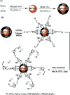
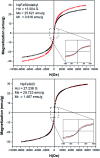
Magnetic nanoparticles with specific surface features are interesting materials for biomedical applications. The combination of molecular interactions on small particles with macroscopic cohesion forces offers unique opportunities. This work reports the synthesis of magnetic core-shell nanoparticles with alkylimidazolium coated surface for effective DNA extraction. A magnetic Fe2O3 core was coated with a silica shell and functionalized with an organic halide. This enabled a surface coating with organic cations to mediate effective molecular interactions with polyanionic DNA. The large surface area of the ∼20 nm small particles with a magnetization of 25 emu g-1 enabled high DNA particle loading of 1/30 m% with easy isolation based on an external magnetic field. Moreover, the coating of the particles stabilized DNA against ultrasound initiated fragmentation.
This journal is © The Royal Society of Chemistry.
Conflict of interest statement
The authors declare no conflict of interest.
Figures





References
-
- Pankhurst Q. A. Connolly J. Jones S. K. Dobson J. J. Phys. D: Appl. Phys. 2003;36:R167.
-
- Halim S. C. Stark W. J. Chimia. 2008;62:13–17. doi: 10.2533/chimia.2008.13. - DOI
-
- Hybrid Nanocomposites for Nanotechnology, ed. L. Merhari, Springer US, New York, 2009
-
- Bodaghifard M. A. Hamidinasab M. Ahadi N. Curr. Org. Chem. 2018;22:234–267. doi: 10.2174/1385272821666170705144854. - DOI
LinkOut - more resources
Full Text Sources

