. 2019 Feb 26;9(12):6643-6649.
doi: 10.1039/c8ra09943j. eCollection 2019 Feb 22.

A colorimetric and ratiometric fluorescent sensor for sequentially detecting Cu2+ and arginine based on a coumarin-rhodamine B derivative and its application for bioimaging

Affiliations

A colorimetric and ratiometric fluorescent sensor for sequentially detecting Cu2+ and arginine based on a coumarin-rhodamine B derivative and its application for bioimaging

Shuai Wang et al. RSC Adv. .

Abstract

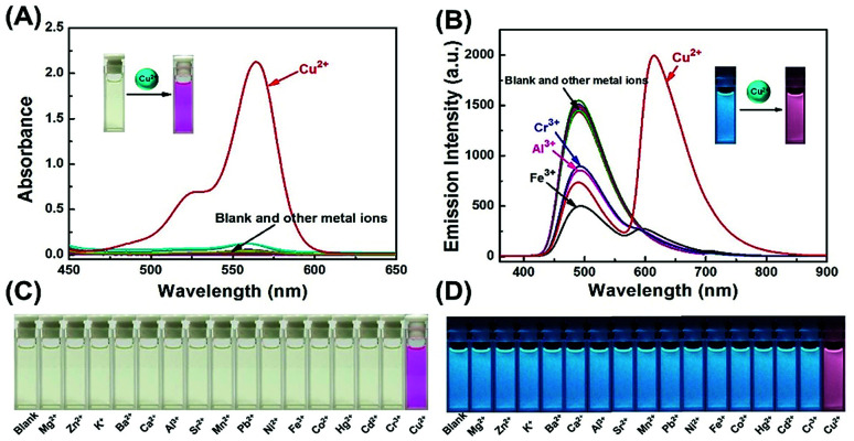
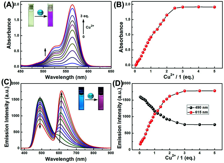
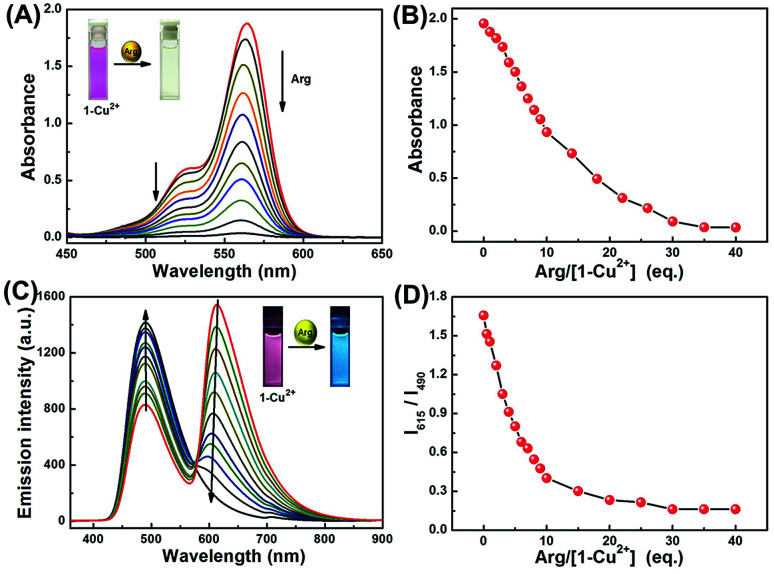
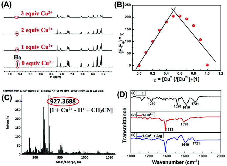
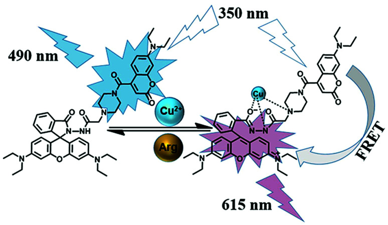
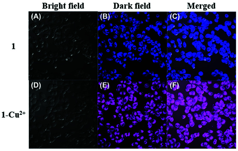
In this work, a colorimetric and ratiometric fluorescent sensor based on a coumarin-rhodamine B hybrid for the sequential recognition of Cu2+ and arginine (Arg) via the FRET mechanism was designed and synthesized. With the addition of Cu2+, the solution displayed a colorimetric change from pale yellow to pink which is discernible by the naked eye. Additionally, the fluorescence intensities of the sensor exhibited ratiometric changes for the detection of Cu2+ at 490 and 615 nm under a single excitation wavelength of 350 nm, which corresponded to the emissions of coumarin and rhodamine B moieties, respectively. The fluorescence color change could be visualized from blue to pink. The limits of detection were determined to be as low as 0.50 and 0.47 μM for UV-vis and fluorescence measurements, respectively. More importantly, the sensor not only can recognize Cu2+ and form a sensor-Cu2+ complex but can also sequentially detect Arg with the resulting complex. The detection limits for Arg were as low as 0.60 μM (UV-vis measurement) and 0.33 μM (fluorescence measurement), respectively. A fluorescence imaging experiment in living cells demonstrated that the fabricated sensor could be utilized in ratiometric fluorescence imaging towards intracellular Cu2+, which is promising for the detection of low-level Cu2+ and Arg with potentially practical significance.


Conflict of interest statement

There are no conflicts to declare.

Figures

Scheme 1
Scheme 1. Structure and synthesis of 1.
Fig. 1
Fig. 1. Spectroscopic changes of 1 (2.0 × 10⁻⁵ mol L−1) induced by the addition of various metal ions (3.0 eq.) in CH3CN–H2O (9/1, v/v) solutions. (A) Absorption spectral changes. (B) Fluorescence emission intensity changes. (C) The color image set upon the addition of various metal ions. (D) The fluorescence image set upon the addition of various metal ions.
Fig. 2
Fig. 2. (A) Changes in absorption spectra of 1 in CH3CN–H2O (9/1, v/v) solutions upon addition of Cu2+. (B) Absorption titration profile (at 564 nm) versus concentration of Cu2+ for 1. (C) Fluorescence spectra of 1 upon titration with Cu2+ (0–5.0 eq.) in CH3CN–H2O (9/1, v/v). Inset: the color change in fluorescence of 1. (D) Fluorescence intensity of sensor 1 at 615 nm (red dot) and 490 nm (black dot) as a function of the Cu2+ concentration (0–5.0 eq.). λex = 350 nm, slit: 5 nm/5 nm.
Fig. 3
Fig. 3. (A) Changes in absorption spectra of 1-Cu2+ in CH3CN–H2O (9/1, v/v) solutions upon addition of Arg (0–40.0 eq.). (B) Absorption titration profile (at 564 nm) versus concentration of Arg for 1-Cu2+. (C) Fluorescent intensities ratio (I615/I490) change of 1-Cu2+ solution in CH3CN–H2O (9/1, v/v, 2 × 10⁻⁵ mol L−1) on incremental addition of Arg (0–40.0 eq.). (D) The relationship between fluorescent intensities ratio (I615/I490) change.
Fig. 4
Fig. 4. (A) Partial 1H NMR spectra change of 1 in DMSO-d6 upon addition of Cu2+. (B) Job's plot for the complex of 1 with Cu2+, indicating the formation of a 1 : 1 complex. The total [1] + [Cu2+] = 40 μM. (C) ESI-MS spectrum of 1-Cu2+. (D) Partial FT-IR spectra of 1 (a), 1-Cu2+ (b) and 1-Cu2+ + Arg (c).
Scheme 2
Scheme 2. Principle for sequentially detecting Cu2+ and arginine.
Fig. 5
Fig. 5. Confocal fluorescence images of HeLa cells incubated with sensor 1 (20 μM) for 30 min (A–C) and then treated with Cu2+ (2 mM) for another 30 min (D–F). Images were obtained using an excitation of 405 nm and emission channels of (B) at 430–530 nm and (E) at 550–650 nm; (C and F) merge images of (A, B and D, E); (A and D) bright field images of the cell culture.

Similar articles

Cited by

References

    1. Que E. L. Domaille D. W. Chang C. J. Chem. Rev. 2008;108:1517–1549. doi: 10.1021/cr078203u. - DOI - PubMed
    1. Wang Y. H. Zhang C. Chen X. C. Yang B. Yang L. Jiang C. L. Nanoscale. 2016;8:5977–5984. doi: 10.1039/C6NR00430J. - DOI - PubMed
    1. Ryu H. Choi M. G. Cho E. J. Chang S. K. Dyes Pigm. 2018;153:117–124. doi: 10.1016/j.dyepig.2018.02.013. - DOI
    1. Jiao Y. Zhou L. He H. Y. Yin J. Q. Gao Q. M. Wei J. N. Talanta. 2018;184:143–148. doi: 10.1016/j.talanta.2018.01.073. - DOI - PubMed
    1. Lan G. Y. Huang C. C. Chang H. T. Chem. Commun. 2010;46:1257–1259. doi: 10.1039/B920783J. - DOI - PubMed