. 2022 Apr 20:16:878154.
doi: 10.3389/fncel.2022.878154. eCollection 2022.

Canonical Bone Morphogenetic Protein Signaling Regulates Expression of Aquaporin-4 and Its Anchoring Complex in Mouse Astrocytes

Affiliations

Canonical Bone Morphogenetic Protein Signaling Regulates Expression of Aquaporin-4 and Its Anchoring Complex in Mouse Astrocytes

Nadia Skauli et al. Front Cell Neurosci. .

Abstract

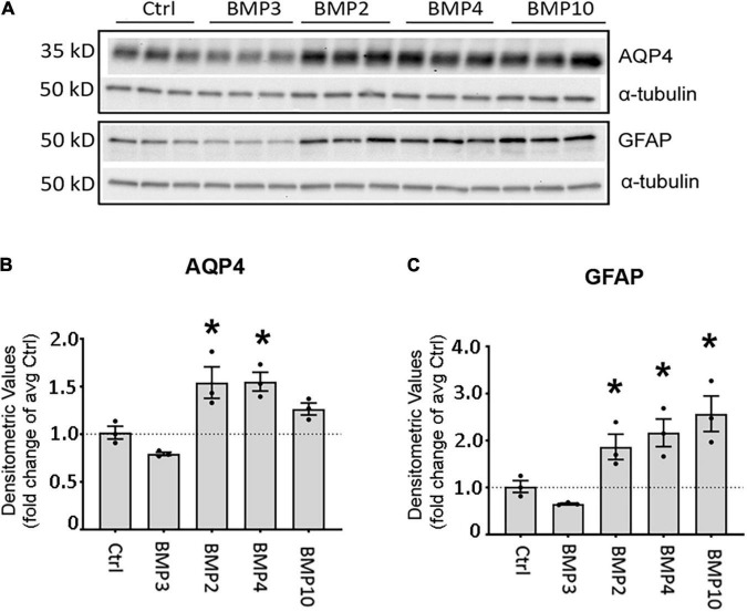
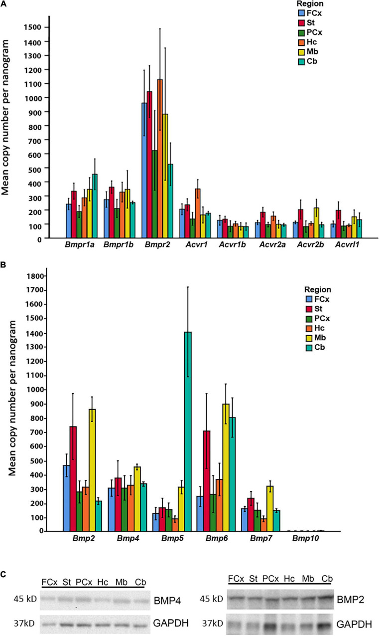
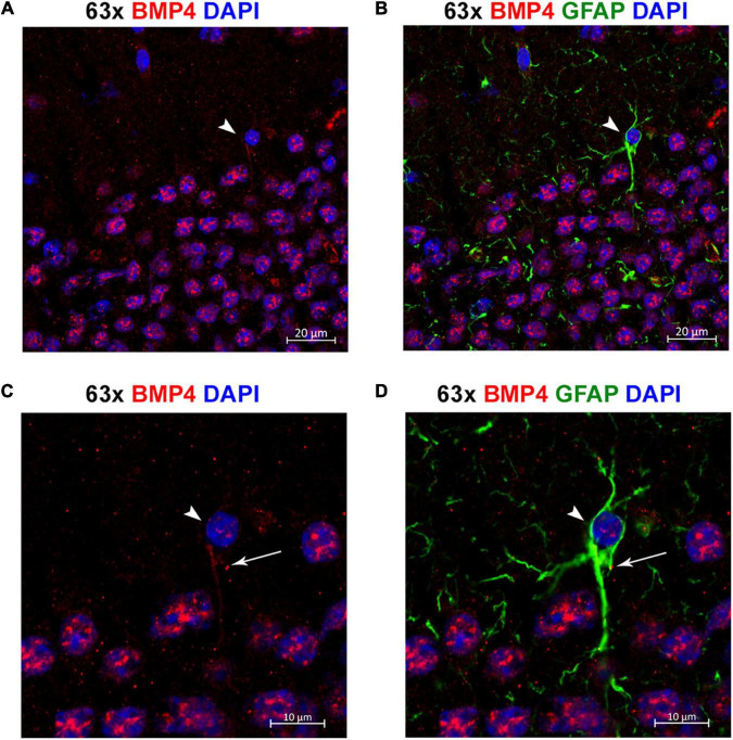
Aquaporin-4 (AQP4) is the predominant water channel in the brain; it is enriched in astrocytic foot processes abutting vessels where it is anchored through an interaction with the dystrophin-associated protein (DAP) complex. Enhanced expression with concomitant mislocalization of AQP4 along astrocyte plasma membranes is a hallmark of several neurological conditions. Thus, there is an urgent need to identify which signaling pathways dictate AQP4 microdistribution. Here we show that canonical bone morphogenetic proteins (BMPs), particularly BMP2 and 4, upregulate AQP4 expression in astrocytes and dysregulate the associated DAP complex by differentially affecting its individual members. We further demonstrate the presence of BMP receptors and Smad1/5/9 pathway activation in BMP treated astrocytes. Our analysis of adult mouse brain reveals BMP2 and 4 in neurons and in a subclass of endothelial cells and activated Smad1/5/9 in astrocytes. We conclude that the canonical BMP-signaling pathway might be responsible for regulating the expression of AQP4 and of DAP complex proteins that govern the subcellular compartmentation of this aquaporin.

Keywords: Smad1/5/9; aquaporin-4; astrocyte; bone morphogenetic protein; dystrophin.


Conflict of interest statement

The authors declare that the research was conducted in the absence of any commercial or financial relationships that could be construed as a potential conflict of interest.

Figures

FIGURE 1
FIGURE 1
BMPs upregulate AQP4 in mESC astrocytes and primary mouse astrocytes after 6 days treatment. (A) Plate Runner assay on mESC derived astrocytes treated with BMPs and immunostained with an antibody to AQP4 demonstrate stronger immunofluorescence intensities (green) in calls treated with BMPs –2, –4, –5, –6, –7, and –10. AQP4 staining shown in green, nuclear staining with DAPI is shown in white pseudocolor. (B) Assessment of fluorescence intensity reveals a significant increase in AQP4 staining in cells treated with BMPs –2, –4, 5, 6, 7, and 10 when comparing mean average cell intensities. The AQP4 immunofluorescent intensity in cells treated with BMP1 and BMP3 is not different from that of the untreated cells (CTRL). Staining intensity is presented as fold changes of mean average cell intensity normalized to that of untreated cells. *Sig ANOVA LSD post-hoc test, horizontal lines with * indicates significance in several samples; error bars: SEM; P < 0.05; n = 4. (C) Plate Runner assay on matured primary astrocytes treated with BMPs and immunostained with antibodies to AQP4 (red) and GFAP (green) demonstrates stronger immunofluorescence intensities in cells treated with BMPs –2, –4, –5, –6, –7, and –10. AQP4 staining shown in red, nuclear staining with DAPI shown in blue, GFAP staining shown in green with 20× and 40× magnification. (D) Assessment of fluorescence intensity in treated primary astrocytes reveals a significant increase in AQP4 and GFAP staining in cells treated with BMPs –2, –4, –5, –6, –7, and –10 when comparing mean average cell intensities. BMP1, BMP3, and FGF2 do not affect AQP4 staining intensities Staining intensity is presented as fold changes of mean average cell intensity normalized to that of untreated cells (CTRL). *Sig ANOVA LSD post-hoc test, horizontal lines with *indicates significance in several samples; error bars: SEM; P < 0.05; n = 4. (E) Representative immunoblot of primary astrocytes treated with BMPs demonstrates that AQP4 expression on the protein level is increased by BMPs –2, –4, –6, –7, and –10, while corresponding α-tubulin expression is not affected. Samples from two separate experiments were run in parallel in the same gel with identical results, n = 2.
FIGURE 2
FIGURE 2
Canonical BMPs transcriptionally regulate Aqp4, Gfap, and DAP complex genes. RT-qPCR of Aqp4 (A), Gfap (B), DAP complex genes Snta1 (C), Dag1 (D), Dtna (E), and Dmd (F); (DP71 transcript variant) on cDNA from BMP treated primary mature astrocytes confirms transcriptional regulation by canonical BMPs. Transcripts differentially regulated by BMPs at 12 and 72 h. Expression is shown as fold changes compared to untreated cells (Ctrl). Red coloring indicates significant upregulation; Blue coloring indicates significant downregulation. *Sig ANOVA LSD post-hoc test; error bars: SEM; dots: individual sample values; P < 0.05; n = 3.
FIGURE 3
FIGURE 3
Canonical BMPs increase expression of AQP4 and GFAP protein in astrocytes. (A) Immunoblot assessment of AQP4 and GFAP protein lysates after treatment with BMPs for 72 h validates upregulation on the protein level only in cells treated with BMPs known to activate Smad1/5/9 (BMPs –2, –4, –10) and not the non-canonical BMP3. (B,C) Densitometric analysis of the immunoblots confirms that both AQP4 and GFAP are significantly upregulated by BMP2, BMP4 and BMP10 treatment. Expression shown as fold changes compared to untreated cells (Ctrl). *Sig ANOVA LSD post-hoc test; error bars: SEM; dots: individual sample values; P < 0.05; n = 3.
FIGURE 4
FIGURE 4
Matured primary astrocytes express BMP receptors and reveal activation of Smad1/5/9 upon treatment with canonical BMPs. (A) RT-qPCR confirms that astrocytes express BMP receptor transcripts. BMP receptor transcripts Bmpr1a, Bmpr1b, Bmpr2, Acvr1, Acvr1b, and Acvr2a are robustly expressed, while Acvr1 exhibits low expression and Acvr1l is not expressed. Values are presented as mean copy numbers per nanogram total RNA input into the cDNA reaction. Dots: individual sample values; Error bars = SEM; n = 4. (B–D) Immunoblots and quantification using antibodies to Smad1 and phosphorylated Smad1/5/9 (pSmad1/5/9) on protein lysates from primary astrocytes after 3 days of BMP treatment. α -tubulin was used as a loading control. *Sig ANOVA LSD post-hoc test; Dots: individual sample values; Error bars = SEM; P < 0.05; n = 3. (C) Smad1 is expressed in all samples and significantly lower in cells treated with BMP2, –4, and –10. (D) pSmad1/5/9 phosphorylation is induced and significantly increased only in samples treated with canonical BMPs –2, –4, and –10.
FIGURE 5
FIGURE 5
Expression of BMP receptors and canonical BMPs in different brain regions. (A) RT-qPCR demonstrates expression of all Bmp receptor transcripts in different regions of adult mouse brain. Bmpr1a and Bmpr1b transcripts are moderately expressed, while Bmpr2 transcripts exhibit particularly high expression in all regions. Acvr1, Acvr1b, Acvr2a, Acvr2b, and Acvrl1 transcripts all show low to moderate expression. Values presented as copy numbers per nanogram total RNA input into the cDNA reaction, Error bars = SEM; n = 4. (B) RT-qPCR demonstrates that all canonical BMPs, with the exception of Bmp10, are expressed in all brain regions investigated. Bmp2 transcripts are expressed throughout the brain, with highest levels in striatum and midbrain. Bmp4 transcripts are moderately expressed throughout all regions. Bmp5 transcripts are moderately expressed throughout the brain, but highly expressed in cerebellum. Bmp6 transcripts are highly expressed in striatum, midbrain and cerebellum, while expression is moderate in other regions. Bmp7 transcripts are moderately expressed in all regions. Bmp10 transcripts were not detected in any of the investigated regions. Values presented as copy numbers per nanogram total RNA input into the cDNA reaction- Error bars = SEM; n = 4. (C) Representative immunoblot shows expression of BMP2 and BMP4 protein in the investigated brain regions in samples from the same animals, n = 4.
FIGURE 6
FIGURE 6
BMP2 and BMP4 are mainly localized in neurons. Confocal images showing BMP2 and BMP4 (in red), the astrocyte marker Glial fibrillary acidic protein (GFAP; in green). Nuclear staining is shown in blue. BMP2 and BMP4 is expressed in neurons in cortex, hippocampus, cerebellum and midbrain. The staining does not co-localize with GFAP in the investigated regions. In midbrain, BMP2 and BMP4 expression is seen in dopaminergic neurons (DAT; white), while BMP4 staining is also observed in vessels. Cx, neocortex; Hc CA, hippocampus CA1 region; Hc DG, hippocampus dentate gyrus; Cb, cerebellum; Mb, midbrain. Scale bars = 50 μm.
FIGURE 7
FIGURE 7
A subpopulation of endothelial cells express BMP2 and BMP4. Confocal images showing BMP2 and BMP4 (green) and the endothelial cell marker tomato lectin (red). Nuclear staining is shown in blue. In some vessels in neocortex, BMP2 staining is observed (arrow), while most vessels are BMP2 negative (asterisks) in neocortex, hippocampus and midbrain. In contrast, widespread BMP4 staining is observed in vessels in cortex, hippocampus and midbrain (arrow). Cx, neocortex; Hc CA, hippocampus CA1 region; Mb, midbrain. Scale bars = 50 μm.
FIGURE 8
FIGURE 8
Individual astrocytes express BMP4. (A,B) Confocal images identifying BMP4 staining in hippocampal astrocytes. Strong BMP4 labeling (red) is localized around the nuclei (blue) of hippocampal neurons, with diffuse and punctate labeling in the parenchyma and weaker labeling in a GFAP (green) positive astrocyte cell body (arrowhead). (C,D) Magnified images focused on the individual astrocyte from (A,B) exhibiting BMP4 labeling mainly localized to the area around the nucleus (arrowhead) with some labeling also present in processes (arrow). Scale bars = 20 μm (A,B), 10 μm (C,D).
FIGURE 9
FIGURE 9
Phosphorylated Smad1/5/9 is mainly localized in subpial and perivascular astrocytes. (A,B) Confocal images, Upper panel with DAPI nuclear staining confirm immunofluorescent phospho-Smad1/5/9 staining in astrocytes. (A) Astrocytes on the cortical surface and in association with blood vessels are positive (arrows) for nuclear phospho-Smad1/5/9 (red), while endothelial cells and pial cells are not. (B) Phospho-Smad1/5/9 (red) staining is present in the nucleus of a GFAP-positive astrocyte associated with a vessels (arrow), but nuclei belonging to endothelial cell lining the vessel, or nuclei in other GFAP-negative cells (some of them with a clear nucleolus) exhibit only very faint or no staining at all. L, vessel lumen. Scale bars = 20 μm.

Similar articles

Cited by

References

    1. Alvestad S., Hammer J., Hoddevik E. H., Skare O., Sonnewald U., Amiry-Moghaddam M., et al. (2013). Mislocalization of AQP4 precedes chronic seizures in the kainate model of temporal lobe epilepsy. Epilepsy Res. 105 30–41. 10.1016/j.eplepsyres.2013.01.006 - DOI - PubMed
    1. Amiry-Moghaddam M., Xue R., Haug F. M., Neely J. D., Bhardwaj A., Agre P., et al. (2004b). Alpha-syntrophin deletion removes the perivascular but not endothelial pool of aquaporin-4 at the blood-brain barrier and delays the development of brain edema in an experimental model of acute hyponatremia. Faseb J. 18 542–544. 10.1096/fj.03-0869fje - DOI - PubMed
    1. Amiry-Moghaddam M., Frydenlund D. S., Ottersen O. P. (2004a). Anchoring of aquaporin-4 in brain: molecular mechanisms and implications for the physiology and pathophysiology of water transport. Neuroscience 129 999–1010. 10.1016/j.neuroscience.2004.08.049 - DOI - PubMed
    1. Amiry-Moghaddam M., Williamson A., Palomba M., Eid T., de Lanerolle N. C., Nagelhus E. A., et al. (2003b). Delayed K+ clearance associated with aquaporin-4 mislocalization: phenotypic defects in brains of alpha-syntrophin-null mice. Proc. Natl. Acad. Sci. U.S.A. 100 13615–13620. 10.1073/pnas.2336064100 - DOI - PMC - PubMed
    1. Amiry-Moghaddam M., Otsuka T., Hurn P. D., Traystman R. J., Haug F. M., Froehner S. C., et al. (2003a). An alpha-syntrophin-dependent pool of AQP4 in astroglial end-feet confers bidirectional water flow between blood and brain. Proc. Natl. Acad. Sci. U.S.A. 100 2106–2111. 10.1073/pnas.0437946100 - DOI - PMC - PubMed

LinkOut - more resources