. 2020 Jun 5;10(36):21399-21405.
doi: 10.1039/d0ra02101f. eCollection 2020 Jun 2.

A pH tuning single fluorescent probe based on naphthalene for dual-analytes (Mg2+ and Al3+) and its application in cell imaging

Affiliations

A pH tuning single fluorescent probe based on naphthalene for dual-analytes (Mg2+ and Al3+) and its application in cell imaging

Chunwei Yu et al. RSC Adv. .

Abstract

In this study, a naphthalene Schiff-base P which serves as a dual-analyte probe for the quantitative detection of Al3+ and Mg2+ has been designed. The proposed probe showed an ''off-on'' fluorescent response toward Al3+ in ethanol-water solution (1 : 9, v/v, pH 6.3, 20 mM HEPES) over other metal ions and anions, while the detection by the probe could be switched to Mg2+ by regulating the pH from 6.3 to 9.4. The sensing mechanisms of P to Al3+/Mg2+ are attributed to inhibition of the photo-induced electron transfer (PET) process by the formation of 1 : 1 ligand-metal complexes. More importantly, the probe was applied successfully in living cells for the fluorescent cell-imaging of Al3+ and Mg2+.


Conflict of interest statement

There are no conflicts to declare.

Figures

Scheme 1
Scheme 1. Synthesis route of probe P.
Fig. 1
Fig. 1. (a) Fluorescence emission spectra of P (10 μM) in response to different metal ions (10 μM) in ethanol–water solution (1 : 9, v/v, 20 mM HEPES, pH 6.3); (b) fluorescence emission spectra of P (10 μM) in response to different metal ions (10 μM) in ethanol–water solution (1 : 9, v/v, 20 mM HEPES, pH 9.4).
Fig. 2
Fig. 2. (a) Fluorescence response of P (10 μM) with various concentrations of Al3+ in ethanol–water solution (1 : 9, v/v, 20 mM HEPES, pH 6.3). Inset: the fluorescence of P (10 μM) as a function of Al3+ concentration (0.5–5.0 μM); (b) fluorescence response of P (10 μM) with various concentrations of Mg2+ in ethanol–water solution (1 : 9, v/v, 20 mM HEPES, pH 9.4). Inset: the fluorescence of P (10 μM) as a function of Mg2+ concentration (1–8.0 μM).
Fig. 3
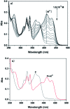
Fig. 3. (a) Absorbance of P (10 μM) with various concentrations of Al3+ in ethanol–water solution (1 : 9, v/v, 20 mM HEPES, pH 6.3); (b) absorbance of P (10 μM) with Al3+ (100 μM) in ethanol–water solution (1 : 9, v/v, 20 mM HEPES, pH 6.3).
Fig. 4
Fig. 4. (a) Absorbance of P (10 μM) with various concentrations of Mg2+ in ethanol–water solution (1 : 9, v/v, 20 mM HEPES, pH 9.4); (b) absorbance of P (10 μM) with Mg2+ (100 μM) in ethanol–water solution (1 : 9, v/v, 20 mM HEPES, pH 9.4).
Fig. 5
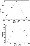
Fig. 5. Job's plot for determining the stoichiometry of P and (a) Al3+ and (b) Mg2+. The total concentration was kept at 50 μM.
Fig. 6
Fig. 6. 1H NMR titration of P with Mg2+.
Scheme 2
Scheme 2. Proposed binding modes of P with Al3+ and Mg2+.
Fig. 7
Fig. 7. (a-I) and (b-I) are the field images of P (10 μM) treated HepG2 cells. (a-II) and (b-II) represent fluorescence images treated with P (10 μM), in the presence of Al3+ or Mg2+ (1 μM), respectively; (a-III) and (b-III) indicate bright field images of cells shown in the panels, respectively. For all imaging, the samples were excited at 375 nm.
Fig. 8
Fig. 8. Confocal fluorescence images of HepG2 cells incubated with P (10 μM) and Hoechst 33 342 (1 μg mL−1) for 30 min. Cells loaded with Al3+ or Mg2+ (10 μM), then treated with P (10 μM) and Hoechst 33 342 (1 μg mL−1) for 30 min. (a-I), (b-I) Green and orange channels with P, respectively; (a-II), (b-II) Blue channel with Hoechst 33 342; (a-III), (b-III) Overlay of images showing fluorescence from P (a-I), (b-I) and Hoechst 33 342 (a-II), (b-II).

References

    1. He X. R. Liu H. B. Li Y. Wang S. Li Y. Wang N. Xiao J. Xu X. Zhu D. Adv. Mater. 2005;17:2811. doi: 10.1002/adma.200501173. - DOI
    2. Quang D. T. Kim J. S. Chem. Rev. 2007;107:3780. doi: 10.1021/cr068046j. - DOI - PubMed
    1. Wang J. L. Lin W. Y. Li W. L. Chem.–Eur. J. 2002;18:13629. doi: 10.1002/chem.201202146. - DOI - PubMed
    2. Schmitz C. Perraud A. Johnson C. O. Inabe K. Smith M. K. Penner R. Kurosaki T. Fleig A. Scharenberg A. M. Cell. 2003;114:191. doi: 10.1016/S0092-8674(03)00556-7. - DOI - PubMed
    1. Stepura O. B. Martynow A. I. Int. J. Cardiol. 2009;134:145. doi: 10.1016/j.ijcard.2009.01.047. - DOI - PubMed
    2. Wolf F. I. Torsello A. Fasanella A. Cittadini A. Mol. Asp. Med. 2003;24:11. doi: 10.1016/S0098-2997(02)00088-2. - DOI - PubMed
    1. Saris N. E. L. Mervaala E. Karppanen H. Khawaja J. A. Lewenstam A. Clin. Chim. Acta. 2000;294:1. doi: 10.1016/S0009-8981(99)00258-2. - DOI - PubMed
    1. Liu Z. C. Yang Z. Y. Li Y. X. Li T. R. Wang B. D. Li Y. Jin X. L. Inorg. Chim. Acta. 2013;395:77. doi: 10.1016/j.ica.2012.10.009. - DOI
    2. Qin J. C. Fan L. Li T. R. Yang Z. Y. Synth. Met. 2015;199:179. doi: 10.1016/j.synthmet.2014.11.030. - DOI
    3. Barceló J. Poschenrieder C. Environ. Exp. Bot. 2002;48:75. doi: 10.1016/S0098-8472(02)00013-8. - DOI
    4. King S. W. Savory J. Willis M. R. Gitelman H. J. Crit. Rev. Clin. Lab. Sci. 1981;14:1. doi: 10.3109/10408368109105861. - DOI - PubMed
    5. Parkinson I. S. Ward M. K. Kerr D. N. J. Clin. Pathol. 1981;34:1285. doi: 10.1136/jcp.34.11.1285. - DOI - PMC - PubMed
    6. Zhou J. Horev B. Hwang G. Klein M. I. Koo H. Benoit D. S. J. Mater. Chem. 2016;4:3075. doi: 10.1039/C5TB02054A. - DOI - PMC - PubMed
    7. Fu Y. L. Tu Y. Y. Fan C. B. Zheng C. H. Liu G. Pu S. Z. New J. Chem. 2016;40:8579. doi: 10.1039/C6NJ01458E. - DOI
    8. Pu S. Z. Zhang C. C. Fan C. B. Liu G. Dyes Pigm. 2016;129:24. doi: 10.1016/j.dyepig.2016.02.001. - DOI
    9. Ding H. C. Li B. Q. Pu S. Z. Liu G. Jia D. C. Yu Z. Sens. Actuators, B. 2017;247:26. doi: 10.1016/j.snb.2017.02.172. - DOI