

The clinical evolution of lichen planus pemphigoides
- PMID: 35518823
- PMCID: PMC9037440
- DOI: 10.1080/08998280.2021.2022065
The clinical evolution of lichen planus pemphigoides
Abstract
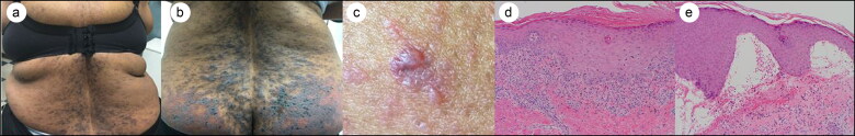
Lichen planus pemphigoides (LPP) is a rare autoimmune subepidermal blistering disease characterized by lichenoid and bullous lesions. LPP is generally thought to be idiopathic, possibly related to medication or malignancy, or potentially the result of long-standing lichenoid inflammation damaging the basement membrane zone leading to epitope spreading. The histological appearance of lichenoid and bullous lesions in LPP resembles findings of lichen planus and bullous pemphigoid, respectively. We present a case of LPP in a 64-year-old woman with a brief historical review of the establishment of LPP as a separate disease entity and a discussion of similarities and differences of LPP with other lichenoid and blistering dermatoses.
Keywords: Blister; bullous; lichen; pemphigoides; planus.
Copyright © 2022 Baylor University Medical Center.
Figures
References
-
- Knisley R, Petropolis A, Mackey V.. Lichen planus pemphigoides treated with ustekinumab. Cutis. 2017;100(6):415–418. - PubMed
-
- Mendiratta V, Asati DP, Koranne RV.. Lichen planus pemphigoides in an Indian female. Indian J Dermatol. 2005;50:224–226.
Publication types
LinkOut - more resources
Full Text Sources