Review
. 2020 Nov 9;10(67):40745-40794.
doi: 10.1039/d0ra08396h.

The oxazolomycin family: a review of current knowledge

Affiliations
Review

The oxazolomycin family: a review of current knowledge

Patrik Oleksak et al. RSC Adv. .

Abstract

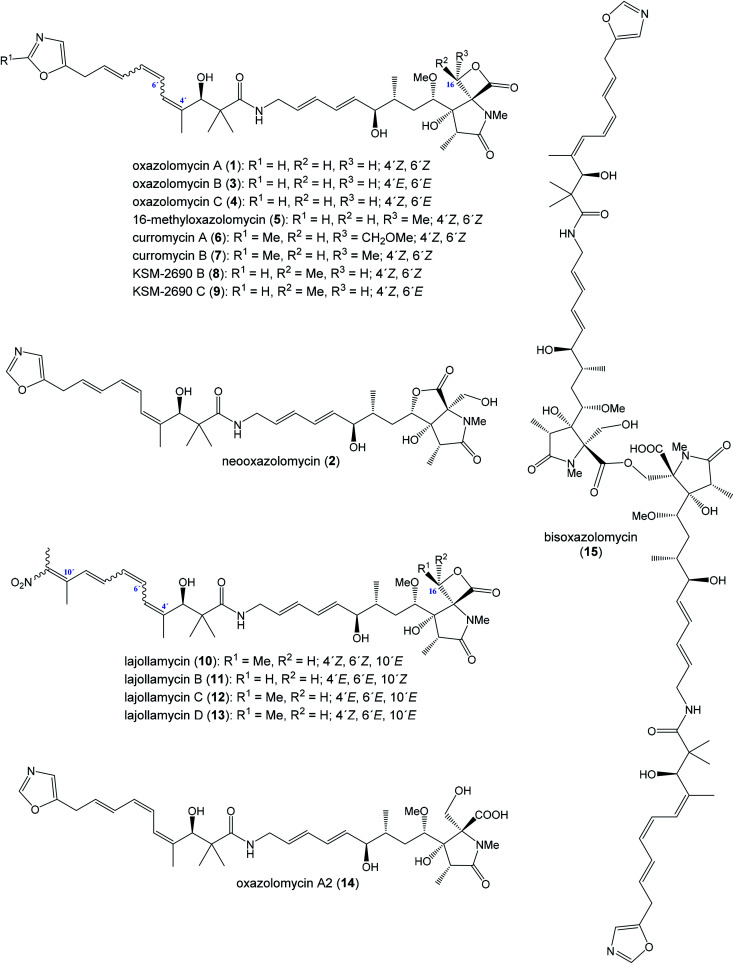
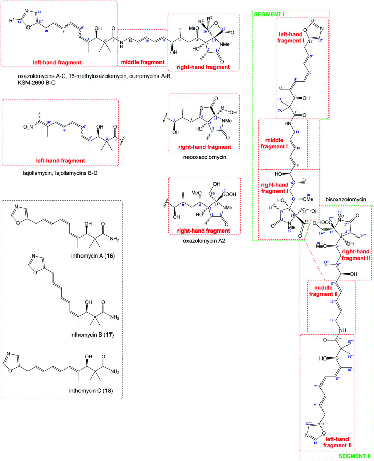
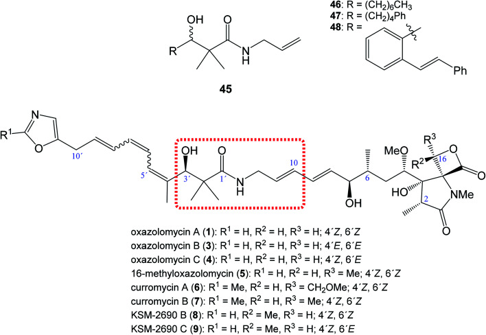
Oxazolomycin A and neooxazolomycin were firstly isolated in 1985 by the group of Uemura et al. from the Streptomyces sp. bacteria. To date, there have been reported 15 different natural compounds commonly classified as part of the oxazolomycin family. All oxazolomycin compounds possess extraordinary structures and they represent a synthetic challenge. Such molecules are additionally known for their wide range of biological activity including antibacterial, antiviral and cytotoxic effects. The present review summarizes the structural elucidation and classification of oxazolomycin compounds, their biosynthesis and biological activity. It is further focused on the total syntheses of oxazolomycins and one formal synthesis reported to date.


Conflict of interest statement

The authors declare that they have no conflict of interest.

Figures

Fig. 1
Fig. 1. Oxazolomycin family members.
Fig. 2
Fig. 2. Structural division of oxazolomycin family members and structure of inthomycins.
Fig. 3
Fig. 3. Genetic organization in the ozm cluster. Numbers refer to genes outside ozm cluster, letters refer to genes inside ozm cluster.
Fig. 4
Fig. 4. Determination of OzmH A domain substrate specificities. The ATP-PPi exchange reactions were performed using amino acids Gly, Ala and Ser as substrates and H2O as a negative control (100% relative activity corresponds to 995 320 cpm).
Fig. 5
Fig. 5. Deletion of ozmM and complementation of the ozmM mutant with either intact ozmM or ozmM-AT1 (Ser81 to Gly) or ozmM-AT2 (Ser402 to Gly) mutant and their effect on the biosynthesis of oxazolomycin A. (A) Schematic representation of constructs for the generation of the ZH9 deletion mutant strain and its genetic complementation strains ZH10, ZH14 and ZH15. Details of side-specific mutagenesis with OzmM-AT1 and OzmM-AT2 are shown. (B) HPLC analysis of oxazolomycin A (1) production in: (I) Streptomyces albus JA3453; (II) ZH9 mutant; (III) ZH10 mutant; (IV) ZH14 mutant and (V) ZH15 mutant.
Fig. 6
Fig. 6. Proposed model of oxazolomycin A biosynthesis in Streptomyces albus JA3453. Used abbreviations: A, adenylation domain; ACP, acyl carrier protein domain; AT1, N-terminal acyl transferase domain of OzmM; AT2, central acyl transferase domain of OzmM; C, condensation domain; DH, dehydratase domain; ER, enoyl reductase domain; F, formylation domain; KR, ketoreductase domain; KS, ketosynthetase domain; MT, methyltransferase domain; OX, oxidoreductase domain; PCP, peptide carrier protein domain; ?, unknown domain; SAM, S-adenosyl methionine.
Scheme 1
Scheme 1. Proposed pathway for methoxymalonyl-ACP extender unit biosynthesis in ozm cluster.
Fig. 7
Fig. 7. Proposed biosynthetic origin of oxazolomycin A (1) according to labeling data and analysis of genes in ozm cluster.
Fig. 8
Fig. 8. Known proteasome inhibitors with pyroglutamate core.
Scheme 2
Scheme 2. Pyroglutamate derivatives and their binding in 20S proteasome.
Fig. 9
Fig. 9. Selected bioactive right-hand fragment derivatives.
Fig. 10
Fig. 10. Position of structural subunit 45 in the structure of oxazolomycins responsible for U-shaped conformation and its derivatives 46–48 prepared by Bagwell et al.
Fig. 11
Fig. 11. Structure and calculated minimum energy conformation of amide 49.
Fig. 12
Fig. 12. Structure and calculated minimum energy conformation of inthomycin B (17).
Fig. 13
Fig. 13. Structure and calculated minimum energy conformation of oxazolomycin B (3).
Fig. 14
Fig. 14. Effects of inthomycin A (16) and inthomycin B (17) on the growth of DU-145 cells alone (black open circles), DU-145 cells cocultured with PrSC (red filled circles) and PrSC alone (blue filled squares) were determined using rhodanile blue staining.
Fig. 15
Fig. 15. Effect of curromycin B (7) and curromycin B diacetate against Agrobacterium tumefaciens growth on an inoculated potato tuber disk. Bacterial cells in the absence (○) and the presence of curromycin B diacetate (●) or curromycin B (Δ) were counted 0, 6, 12, 24 and 48 h after inoculation.
Fig. 16
Fig. 16. Comparison of antibacterial activity of oxazolomycins 1, 14 and 15 using disc diffusion method at the concentration 30 μg per disc.
Fig. 17
Fig. 17. Effect of curromycin A (6), B (7) and azidothymidine (AZT) on primary infected human lymphoid cells. Green columns: % inhibition of reverse transcriptase; black columns: % inhibition of MTT.
Fig. 18
Fig. 18. Effect of curromycin A (6), B (7) and azidothymidine (AZT) in chronic replication assay. Green columns: % inhibition of reverse transcriptase; black columns: % inhibition of MTT.
Fig. 19
Fig. 19. Cytotoxic activity of curromycin A (6), B (7) and 16-methyloxazolomycin (5) against mouse leukemia P388.
Fig. 20
Fig. 20. Comparison of IC50 values for oxazolomycin A (1), A2 (14) and bisoxazolomycin (15) against human leukemia HL60.
Fig. 21
Fig. 21. Inhibition activity Candida albicans isocitrate lyase by lajollamycins 10–13.
Fig. 22
Fig. 22. Effect of curromycin A (6) on the luciferase expression in HT1080 G-L cells. HT1080 G-L cells were treated with 6 in the presence (○) or absence (●) of 2-deoxyglucose (10 mM) for 18 h at 37 °C. The relative luciferase activity compared with non-treated control was measured with a luminometer.
Fig. 23
Fig. 23. Effect on crown gall formation of oxazolomycin A (1) administered simultaneously with the inoculation of Agrobacterium tumefaciens.
Fig. 24
Fig. 24. Growth curves of crown gall tissue on solid Murashige–Skoog medium. Oxazolomycin A (○) at 100 μg ml−1; carbenicillin (▲) at 1000 μg ml−1.
Fig. 25
Fig. 25. Effect of oxazolomycins and curomycins against crown gall formation.
Fig. 26
Fig. 26. Decay of oxazolomycin A (●) and oxazolomycin A dipropionate (○) in the process of crown gall formation. The compounds were administered to potato tuber discs inoculated with A. tumefaciens (A), to potato tuber discs (B) or to a culture of A. tumefaciens (C).
Fig. 27
Fig. 27. Summary of reported phytotoxic activity of oxazolomycins.
Fig. 28
Fig. 28. Tolerated, toxic and lethal doses of oxazolomycin A (1) and curromycins A (6) and B (7).
Scheme 3
Scheme 3. Synthesis of Stille cross coupling partner 63 for left-hand fragment of neooxazolomycin by Kende.
Scheme 4
Scheme 4. Synthesis of the left-hand fragment derivative 72 of neooxazolomycin by Kende.
Scheme 5
Scheme 5. Synthesis of the right-hand and middle fragment derivative 91 of neooxazolomycin by Kende.
Scheme 6
Scheme 6. Finalization of the total synthesis of neooxazolomycin (2) by Kende.
Scheme 7
Scheme 7. Synthesis of the left-hand fragment derivative 72 of neooxazolomycin by Hatakeyama.
Scheme 8
Scheme 8. Synthesis of the right-hand and middle fragment derivative 91 of neooxazolomycin by Hatakeyama.
Scheme 9
Scheme 9. Finalization of the total synthesis of neooxazolomycin (2) by Hatakeyama.
Scheme 10
Scheme 10. Synthesis of the left-hand fragment derivative 132 of neooxazolomycin by Kim.
Scheme 11
Scheme 11. Synthesis of the right-hand and middle fragment derivative 157 of neooxazolomycin by Kim.
Scheme 12
Scheme 12. Finalization of the total synthesis of neooxazolomycin (2) by Kim.
Scheme 13
Scheme 13. Synthesis of the left-hand fragment derivative 72 of oxazolomycin A by Hatakeyama.
Scheme 14
Scheme 14. Synthesis of the right-hand fragment derivative 176 of oxazolomycin A by Hatakeyama.
Scheme 15
Scheme 15. Synthesis of the middle fragment derivative 117 of oxazolomycin A by Hatakeyama.
Scheme 16
Scheme 16. Finalization of the total synthesis of oxazolomycin A (1) by Hatakeyama.
Scheme 17
Scheme 17. Synthesis of the left-hand fragment precursor 188 of lajollamycin B by Hatakeyama.
Scheme 18
Scheme 18. Synthesis of the right-hand and middle fragment derivative 194 of lajollamycin B by Hatakeyama.
Scheme 19
Scheme 19. Hatakeyama's model study for the final Stille coupling in the synthesis of lajollamycin B.
Scheme 20
Scheme 20. Finalization of the total synthesis of lajollamycin B (11) by Hatakeyama.
Fig. 29
Fig. 29. NOESY correlations reported by Hatakeyama et al. for final products in the total synthesis of lajollamycin B.
Scheme 21
Scheme 21. Synthesis of the middle fragment derivative 201 of neooxazolomycin by Taylor.
Scheme 22
Scheme 22. Synthesis of bicyclic precursor for right-hand fragment 208 of neooxazolomycin by Taylor.
Scheme 23
Scheme 23. Synthesis of Stille cross coupling partner 217 for right-hand fragment of neooxazolomycin by Taylor.
Scheme 24
Scheme 24. Synthesis of the right-hand fragment derivative 230 of neooxazolomycin by Taylor.
Fig. 30
Fig. 30. Model for dihydroxylation reported by Taylor et al.
Scheme 25
Scheme 25. Finalization of the formal synthesis of neooxazolomycin (2) by Taylor.
Fig. 31
Fig. 31. Used starting materials for total and formal syntheses of oxazolomycin family members.

Similar articles

Cited by

References

    1. Mori T. Takahashi K. Kashiwabara M. Uemura D. Katayama C. Iwadare S. Shizuri Y. Mitomo R. Nakano F. Matsuzaki A. Tetrahedron Lett. 1985;26:1073–1076. doi: 10.1016/S0040-4039(00)98515-0. - DOI
    1. Takahashi K. Kawabata M. Uemura D. Iwadare S. Mitomo R. Nakano F. Matsuzaki A. Tetrahedron Lett. 1985;26:1077–1078. doi: 10.1016/S0040-4039(00)98516-2. - DOI
    1. Ogura M. Nakayama H. Furihata K. Shimazu A. Seto H. Otake N. J. Antibiot. 1985;38:669–673. doi: 10.7164/antibiotics.38.669. - DOI - PubMed
    1. Ikeda Y. Kondo S. Naganawa H. Hattori S. Hamada M. Takeuchi T. J. Antibiot. 1991;44:453–455. doi: 10.7164/antibiotics.44.453. - DOI - PubMed
    1. Ryu G. Hwang S. Kim S.-K. J. Antibiot. 1997;50:1064–1066. doi: 10.7164/antibiotics.50.1064. - DOI - PubMed

LinkOut - more resources