. 2022 Mar 16;13(4):2174-2185.
doi: 10.1364/BOE.451878. eCollection 2022 Apr 1.

Intraocular scatter compensation with spatial light amplitude modulation for improved vision in simulated cataractous eyes

Affiliations

Intraocular scatter compensation with spatial light amplitude modulation for improved vision in simulated cataractous eyes

Spozmai Panezai et al. Biomed Opt Express. .

Abstract

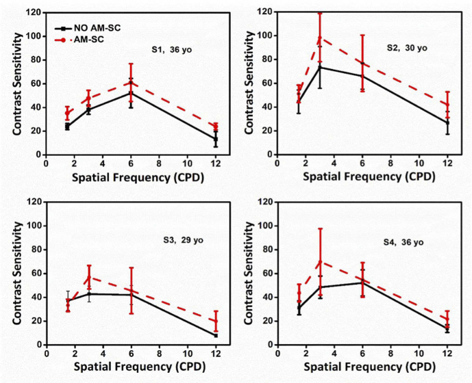
Cataract is one of the common causes of visual impairment due to opacification of the crystalline lens. Increased intraocular scattering affects the vision of cataract patients by reducing the quality of the retinal image. In this study, an amplitude modulation-based scatter compensation (AM-SC) method is developed to minimize the impact of straylight on the retinal image. The performance of the AM-SC method was quantified by numerical simulations of point spread function and retinal images in the presence of different amounts of straylight. The approach was also experimentally realized in a single-pass system with a digital micro-mirror device used as a spatial amplitude modulator. We showed that the AM-SC method allows to enhance contrast sensitivity in the human eyes in vivo with induced scattering.


Conflict of interest statement

The authors declare no conflicts of interest.

Figures

Fig. 1.
Fig. 1.
Concept of the proposed amplitude modulation-based scatter compensation method in cataract eyes. (a) Opacification in the anterior segment of the eye results in reduced retinal image quality (blur, poor contrast). (b) Mapping intraocular light scattering allows for generation of the opacity map of the crystalline lens, which is later transformed into a binary mask. The light entering the eye is spatially modulated by a digital micro-mirror device (DMD) to allow the light to pass through non-opacified regions, which reduces the impact of intraocular scatter on vision.
Fig. 2.
Fig. 2.
(a) Experimental setup implementing AM-SC method equipped with a channel for contrast sensitivity testing. DMD – digital micro-mirror device, L1-L4 – lenses, P – polarizer and A – analyzer. (b) Photograph of the instrument.
Fig. 3.
Fig. 3.
Simulation of impact of intraocular scatter and AM-SC method on the retinal image in the cataract eye: (a) Opacity maps and corresponding binary masks for Log10(s) = 1.75 and Log10(s) =  2.37. (b) PSF with no compensation (PSF No AM-SC) and PSF after scatter compensation (PSF After AM-SC) with AM-SC method. Bar indicates 48 arcminutes. The normalized central intensity profiles along red dashed line plotted for before (No AM-SC; black) and after applying AM-SC method (After AM-SC; red). (c) Ground truth (original) image, retinal blurred image due to the simulated intraocular scattering Log10(s) =  2.37 and the retinal image after applying AM-SC method.
Fig. 4.
Fig. 4.
Retinal image contrast (RIC) for sine-wave gratings of different spatial frequencies and contrast. Relative contrast difference (RCD) between retinal images after implementation of the AM-SC approach. Two levels of straylight parameter were used in calculations: (a), Log10(s) = 1.75, and (b) Log10(s)    2. 37.
Fig. 5.
Fig. 5.
Contrast sensitivity functions (CSFs) measured in four subjects (S1-S4) wearing the spectacles with cataract phantom. Black solid curves represent CFSs with no scatter compensation and red dashed curves represent CSFs after implementation of AM-SC method.

References

    1. Ljungberg M., Pretorius P. H., “SPECT/CT: an update on technological developments and clinical applications,” Br. J. Radiol. 91(1081), 20160402 (2018).10.1259/bjr.20160402 - DOI - PMC - PubMed
    1. Watson C. C., “New, faster, image-based scatter correction for 3D PET,” IEEE Trans. Nucl. Sci. 47(4), 1587–1594 (2000).10.1109/23.873020 - DOI
    1. Horstmeyer R., Ruan H., Yang C., “Guidestar-assisted wavefront-shaping methods for focusing light into biological tissue,” Nat. Photonics 9(9), 563–571 (2015).10.1038/nphoton.2015.140 - DOI - PMC - PubMed
    1. Lysakovski P., Ferrari A., Tessonnier T., Besuglow J., Kopp B., Mein S., Haberer T., Debus J., Mairani A., “Development and benchmarking of a Monte Carlo dose engine for proton radiation therapy,” Front. Phys. 9, 741453 (2021).10.3389/fphy.2021.741453 - DOI
    1. May M. A., Barre N., Kummer K. K., Kress M., Ritsch-Marte M., Jesacher A., “Fast holographic scattering compensation for deep tissue biological imaging,” Nat. Commun. 12(1), 4340 (2021).10.1038/s41467-021-24666-9 - DOI - PMC - PubMed

LinkOut - more resources