. 2022 Mar 4;13(4):1869-1887.
doi: 10.1364/BOE.448038. eCollection 2022 Apr 1.

Lock-in functional near-infrared spectroscopy for measurement of the haemodynamic brain response

Affiliations

Lock-in functional near-infrared spectroscopy for measurement of the haemodynamic brain response

Stanislaw Wojtkiewicz et al. Biomed Opt Express. .

Abstract

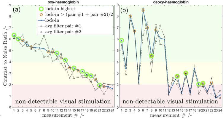
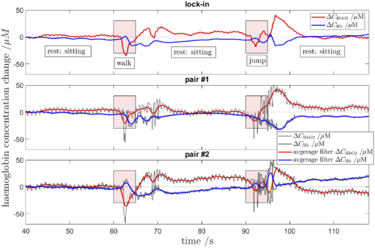
Here we show a method of the lock-in amplifying near-infrared signals originating within a human brain. It implies using two 90-degree rotated source-detector pairs fixed on a head surface. Both pairs have a joint sensitivity region located towards the brain. A direct application of the lock-in technique on both signals results in amplifying common frequency components, e.g. related to brain cortex stimulation and attenuating the rest, including all components not related to the stimulation: e.g. pulse, instrumental and biological noise or movement artefacts. This is a self-driven method as no prior assumptions are needed and the noise model is provided by the interfering signals themselves. We show the theory (classical modified Beer-Lambert law and diffuse optical tomography approaches), the algorithm implementation and tests on a finite element mathematical model and in-vivo on healthy volunteers during visual cortex stimulation. The proposed hardware and algorithm complexity suit the entire spectrum of (continuous wave, frequency domain, time-resolved) near-infrared spectroscopy systems featuring real-time, direct, robust and low-noise brain activity registration tool. As such, this can be of special interest in optical brain computer interfaces and high reliability/stability monitors of tissue oxygenation.


Conflict of interest statement

The authors declare no conflicts of interest related to this article.

Figures

Fig. 1.
Fig. 1.
Sensitivity volumes of light attenuation measurements to changes in absorption for the proposed geometry of two 90-degree rotated source-detector pairs (30 mm source-detector distance). Results of FEM analysis (details of the model presented in method section) for a two-layer model where the upper layer is 13 mm thick. Sensitivities (mean partial path lengths) for pair #1 (a) and pair #2 (b), the sum of both (c) and the square root of the element-wise product (d) are within the same colour scale.
Fig. 2.
Fig. 2.
FEM model used for testing the lock-in signal processing principle (a). Combinations of signals to lock-in (b). ‘long’ refers to 30 mm source-detector separation and ‘short’ is 15 mm. Superficial layer thickness is 13 mm.
Fig. 3.
Fig. 3.
Finite elements 2-layer head model haemodynamic parameters. Biological noise in the extracerebral tissue as measured in-vivo in this study at 15 mm source-detector separation.
Fig. 4.
Fig. 4.
Sources and detectors arranged within a mesh (a) fixed on a head surface (b) over the left side of visual cortex of a healthy volunteer. Panel (a) shows all possible combinations of pairs.
Fig. 5.
Fig. 5.
Data lock-in amplification strategies: (a) the lock-in method – both pairs at large distance (30 mm) with overlapping sensitivity region, (b) both pairs at large distance but no overlapping sensitivity region, (c) collinear pairs – one pair is at large distance and the second one is at the short one (15 mm), (d) both pairs are at the short distance.
Fig. 6.
Fig. 6.
Time courses of ground truth changes in haemoglobin concentrations (thick lines) and results of recovery for overlapping and non-overlapping geometries. The top row of the time series shows results of the typical modified Beer-Lambert law analysis at long (30 mm) and short (15 mm) source-detector distances. The light attenuation panel shows the entry data for the analysis. The amp. error represents the amplitude fit error in percent and the off. error shows error in offset between both periodic signals. Data for the short distance does not provide the brain haemodynamic response and the recovered haemoglobin changes approach zero. The bottom row of panels shows combinations of lock-in filtered data as proposed in Fig. 2(b).
Fig. 7.
Fig. 7.
The frequency analysis of simulated temporal data from Fig. 6. Comparison of the lock-in and the classical data processing approach. Please see the removal of the DC component and frequencies around the visual stimulation using the lock-in filter.
Fig. 8.
Fig. 8.
Good quality data example. Time courses of changes in concentrations of oxy- (red lines) and deoxy-haemoglobin (blue lines) registered in-vivo in a healthy volunteer under visual stimulation for varying lock-in signal strategies (see Fig. 4 and 5). Results for pair #1 and #2 show the average filtering (thick lines) overlaying direct photodetector signals (thin black/grey lines). For high-resolution see the supplemental material.
Fig. 9.
Fig. 9.
Example of a less pronounce visual response. Time courses of changes in concentrations of oxy- (red lines) and deoxy-haemoglobin (blue lines) registered in-vivo in a healthy volunteer under visual stimulation for varying lock-in signal strategies (see Fig. 4 and 5). Results for pair #1 and #2 show the average filtering (thick lines) overlaying direct photodetector signals (thin black/grey lines). For high-resolution see the supplemental material.
Fig. 10.
Fig. 10.
An example of the lock-in method frequency characteristics as compared to a common moving average filtering. The moving average filtering is a zero-phase filter (MATLAB ‘filtfilt’ function). Time data shown in the first column in Fig. 8 (quality data) were used. The oxygenated haemoglobin spectrum is shown.
Fig. 11.
Fig. 11.
The lock-in method with large lock-in sliding window size. Data equal to the first column in Fig. 8 (quality data).
Fig. 12.
Fig. 12.
The CNR in oxygenated (a) and deoxygenated (b) haemoglobin for six subjects and four 90-degree shifted lock-in pairs per subject (a total of 24 measurements). Results are sorted by the CNR of the oxygenated haemoglobin for the lock-in method. Measurements where the lock-in method provides the CNR gain are highlighted with circle markers (big green and smaller orange). Three quality zones in CNR are marked with colours. The bottom one (CNR < 2) marks measurements with non-detectable brain haemodynamic response to the visual stimuli.
Fig. 13.
Fig. 13.
Results of the in-vivo motion artefacts testing. First 40 s of flat rest signal not shown to zoom in into the motion artefact region.

Similar articles

Cited by

References

    1. Xu X., Liu H., Wang L. V., “Time-reversed ultrasonically encoded optical focusing into scattering media,” Nat. Photonics 5(3), 154–157 (2011).10.1038/nphoton.2010.306 - DOI - PMC - PubMed
    1. Caldwell M., Scholkmann F., Wolf U., Wolf M., Elwell C., Tachtsidis I., “Modelling confounding effects from extracerebral contamination and systemic factors on functional near-infrared spectroscopy,” NeuroImage 143, 91–105 (2016).10.1016/j.neuroimage.2016.08.058 - DOI - PMC - PubMed
    1. Pfeifer M. D., Scholkmann F., Labruyère R., “Signal Processing in Functional Near-Infrared Spectroscopy (fNIRS): Methodological Differences Lead to Different Statistical Results,” Front. Hum. Neurosci. 11, 641 (2018).10.3389/fnhum.2017.00641 - DOI - PMC - PubMed
    1. Milej D., Abdalmalak A., Rajaram A., St. Lawrence K., “Direct assessment of extracerebral signal contamination on optical measurements of cerebral blood flow, oxygenation, and metabolism,” Neurophotonics 7(04), 045002 (2020).10.1117/1.NPh.7.4.045002 - DOI - PMC - PubMed
    1. Tachtsidis I., Scholkmann F., “False positives and false negatives in functional near-infrared spectroscopy: issues, challenges, and the way forward,” Neurophotonics 3(3), 031405 (2016).10.1117/1.NPh.3.3.031405 - DOI - PMC - PubMed

LinkOut - more resources