

Decolorization and degradation analysis of Disperse Red 3B by a consortium of the fungus Aspergillus sp. XJ-2 and the microalgae Chlorella sorokiniana XJK
- PMID: 35519313
- PMCID: PMC9064126
- DOI: 10.1039/c9ra01169b
Decolorization and degradation analysis of Disperse Red 3B by a consortium of the fungus Aspergillus sp. XJ-2 and the microalgae Chlorella sorokiniana XJK
Abstract
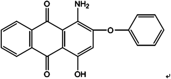
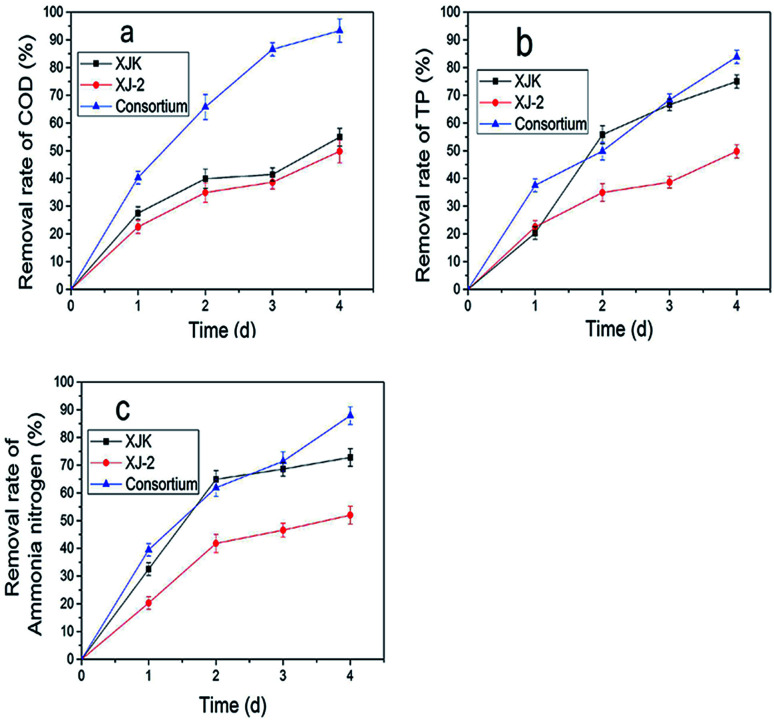
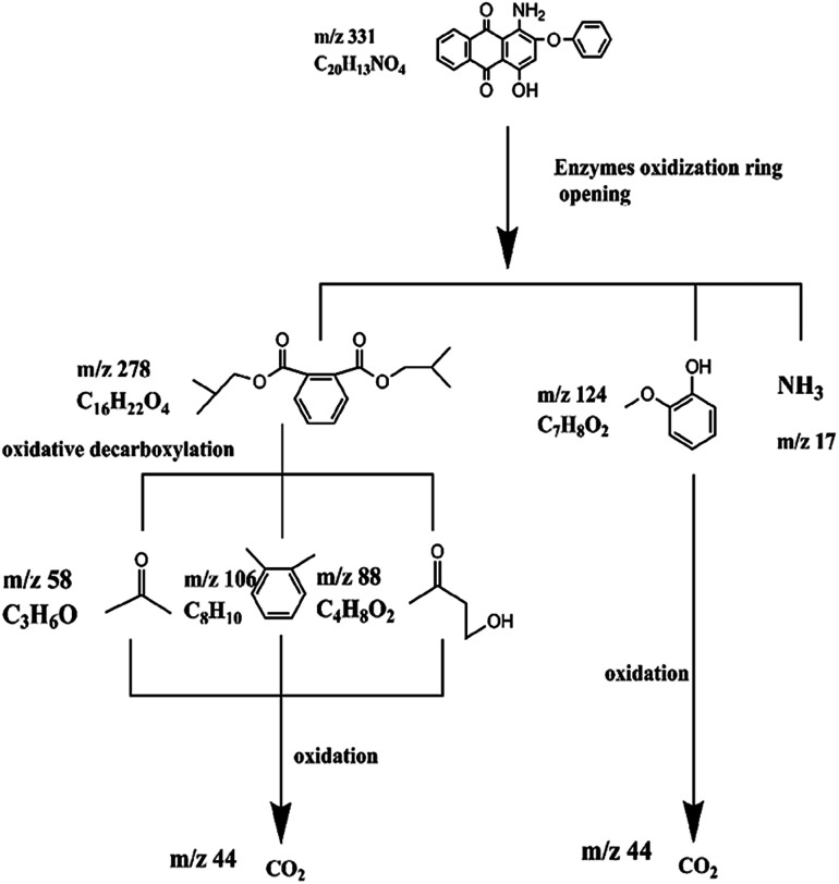
Disperse Red 3B, an anthraquinone dye, was decolorized by a consortium, which was constituted of the fungus (Aspergillus sp. XJ-2) and the microalgae (Chlorella sorokiniana XJK). The consortium performed better than the single system in terms of decolorization and nutrient removal simultaneously in the simulated wastewater of Dispersed Red 3B. The decolorization rate could reach 98.09% by the consortium under the optimized conditions. The removal rate of COD (Chemical Oxygen Demand), TP (Total Phosphorus), and ammonia nitrogen reached 93.9%, 83.9% and 87.6%. Also, the consortium could tolerate higher salt and dye concentration than the single system did. In this co-cultural system, the lignin peroxidase and manganese peroxidase enzyme activities contributed to the degradation of Disperse Red 3B, which reached 86.7 U L-1 and 122.5 U L-1. The result of fermentation liquid analysis with UV-vis, FTIR and GC-MS showed that the colored functional group of the dye was broken and the Dispersed Red 3B was degraded into small molecular compounds with low toxicity. It was suggested that degradation plays a major role during the color removal process. The consortium exhibited greater potential in terms of color removal and water pollutant removal than the separate system did.
This journal is © The Royal Society of Chemistry.
Conflict of interest statement
There are no conflicts to declare.
Figures


Similar articles
-
Decolorization pathways of anthraquinone dye Disperse Blue 2BLN by Aspergillus sp. XJ-2 CGMCC12963.Bioengineered. 2017 Sep 3;8(5):630-641. doi: 10.1080/21655979.2017.1300728. Epub 2017 Mar 8. Bioengineered. 2017. PMID: 28272975 Free PMC article.
-
Biodegradation of a monochlorotriazine dye, cibacron brilliant red 3B-A in solid state fermentation by wood-rot fungal consortium, Daldinia concentrica and Xylaria polymorpha: Co-biomass decolorization of cibacron brilliant red 3B-A dye.Int J Biol Macromol. 2018 Dec;120(Pt A):19-27. doi: 10.1016/j.ijbiomac.2018.08.068. Epub 2018 Aug 15. Int J Biol Macromol. 2018. PMID: 30118766
-
Growth rate, organic carbon and nutrient removal rates of Chlorella sorokiniana in autotrophic, heterotrophic and mixotrophic conditions.Bioresour Technol. 2013 Sep;144:8-13. doi: 10.1016/j.biortech.2013.06.068. Epub 2013 Jun 27. Bioresour Technol. 2013. PMID: 23850820
-
Efficient biodegradation of Congo red dye using fungal consortium incorporated with Penicillium oxalicum and Aspergillus tubingensis.Folia Microbiol (Praha). 2022 Feb;67(1):33-43. doi: 10.1007/s12223-021-00915-8. Epub 2021 Sep 1. Folia Microbiol (Praha). 2022. PMID: 34468947
-
Development of a bioreactor for remediation of textile effluent and dye mixture: a plant-bacterial synergistic strategy.Water Res. 2013 Mar 1;47(3):1035-48. doi: 10.1016/j.watres.2012.11.007. Epub 2012 Nov 23. Water Res. 2013. PMID: 23245543
Cited by
-
Using Fungi in Artificial Microbial Consortia to Solve Bioremediation Problems.Microorganisms. 2024 Feb 26;12(3):470. doi: 10.3390/microorganisms12030470. Microorganisms. 2024. PMID: 38543521 Free PMC article. Review.
-
Statistical modeling of methylene blue degradation by yeast-bacteria consortium; optimization via agro-industrial waste, immobilization and application in real effluents.Microb Cell Fact. 2021 Dec 30;20(1):234. doi: 10.1186/s12934-021-01730-z. Microb Cell Fact. 2021. PMID: 34965861 Free PMC article.
-
Methyl Orange Biodegradation by Immobilized Consortium Microspheres: Experimental Design Approach, Toxicity Study and Bioaugmentation Potential.Biology (Basel). 2022 Jan 5;11(1):76. doi: 10.3390/biology11010076. Biology (Basel). 2022. PMID: 35053074 Free PMC article.
-
Iron-dependent mutualism between Chlorella sorokiniana and Ralstonia pickettii forms the basis for a sustainable bioremediation system.ISME Commun. 2022 Sep 15;2(1):83. doi: 10.1038/s43705-022-00161-0. eCollection 2022. ISME Commun. 2022. PMID: 36407791 Free PMC article.
-
Recent advances in biodecolorization and biodegradation of environmental threatening textile finishing dyes.3 Biotech. 2022 Sep;12(9):186. doi: 10.1007/s13205-022-03247-7. Epub 2022 Jul 21. 3 Biotech. 2022. PMID: 35875175 Free PMC article. Review.
References
-
- Popli S. Patel U. D. Int. J. Environ. Sci. Technol. 2015;12:405–420. doi: 10.1007/s13762-014-0499-x. - DOI
-
- Si J. Cui B.-K. Dai Y.-C. Ann. Microbiol. 2013;63:1099–1108. doi: 10.1007/s13213-012-0567-8. - DOI
-
- Hadibarata T. Yusoff A. R. M. Kristanti R. A. Water, Air, Soil Pollut. 2012;223:933–941. doi: 10.1007/s11270-011-0914-6. - DOI
LinkOut - more resources
Full Text Sources
Miscellaneous