Review
. 2019 Jun 24;9(34):19571-19583.
doi: 10.1039/c9ra02985k. eCollection 2019 Jun 19.

Next-generation of selective histone deacetylase inhibitors

Affiliations
Review

Next-generation of selective histone deacetylase inhibitors

Feifei Yang et al. RSC Adv. .

Abstract

Histone deacetylases (HDACs) are clinically validated epigenetic drug targets for cancer treatment. HDACs inhibitors (HDACis) have been successfully applied against a series of cancers. First-generation inhibitors are mainly pan-HDACis that target multiple isoforms which might lead to serious side effects. At present, the next-generation HDACis are mainly focused on being class- or isoform-selective which can provide improved risk-benefit profiles compared to non-selective inhibitors. Because of the rapid development in next-generation HDACis, it is necessary to have an updated and state-of-the-art overview. Here, we summarize the strategies and achievements of the selective HDACis.


Conflict of interest statement

The authors declare no conflict of interest.

Figures

Fig. 1
Fig. 1. Representative class I HDAC complexes.
Fig. 2
Fig. 2. The common pharmacophore model of HDACis. (A) Space-filling representation of TSA in the active-site pocket of HDLP. (B) Schematic representation of HDLP–TSA interactions (adapted with permission from ). (C) Crystal structure of HDAC8 with SAHA. (D) Binding mode of SAHA with HDAC8 (this figure was cited from ref. 63).
Fig. 3
Fig. 3. Examples of HDACis with different ZBG types.
Fig. 4
Fig. 4. (A) Class I HDAC crystal structures (this figure was cited from ref. 79). (B) Surface representation of the pocket accommodating the linker and capping groups of the HDAC8-selective inhibitors. (C) Surface representation of the same region in hHDAC3 (this figure was cited from ref. 80).
Fig. 5
Fig. 5. Structures of some benzamides selective inhibitors.
Fig. 6
Fig. 6. Structures of some benzamides selective inhibitors.
Fig. 7
Fig. 7. Chemical structure of compound 20 (PCI34051) and the docking pose of PCI34051 in the side pocket of HDAC8 (PDB ID 2VX5) (this figure was cited from ref. 90).
Fig. 8
Fig. 8. Structures of tetrahydroisoquinoline-based HDAC8 inhibitors.
Fig. 9
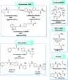
Fig. 9. Structures of novel HDAC8 inhibitors.
Fig. 10
Fig. 10. Structures of pyrazole-based HDAC8 inhibitors.
Fig. 11
Fig. 11. Structures of heteroaryl-based HDAC8 inhibitors.
Fig. 12
Fig. 12. Structures of triazole-based HDAC8 inhibitors.
Fig. 13
Fig. 13. Structures of triazole-based HDAC8 inhibitors.
Fig. 14
Fig. 14. Structures of ortho-aryl N-hydroxycinnamide-based HDAC8 inhibitors.
Fig. 15
Fig. 15. Structures of tetrahydroisoquinoline-based HDAC8 inhibitors.
Fig. 16
Fig. 16. (A) HDAC6 harbors a unique substrate-binding site (this figure was cited from ref. 104). (B) Superposition of broad-specificity HDACis (thin wheat stick figures) and the HDAC6 selective inhibitor HPOB (thin orange stick figure). (C) Close-up view of the Zn2+ binding site in the hCD2 complexes with broad-specificity HDACis. (D) Close-up view of the Zn2+ binding site in the hCD2-HPOB complex (this figure was cited from ref. 19).
Fig. 17
Fig. 17. Structures of novel HDAC6 inhibitors.
Fig. 18
Fig. 18. Structures of heteroaryl-based HDAC6 inhibitors.
Fig. 19
Fig. 19. Structures of quinazoline-based HDAC6 inhibitors.
Fig. 20
Fig. 20. Structures of novel HDAC6 inhibitors.
Fig. 21
Fig. 21. Structures of pyrrolopyrimidine based HDAC6 inhibitors.
Fig. 22
Fig. 22. Structures of carboline based HDAC6 inhibitors.
Fig. 23
Fig. 23. Structures of carboline based HDAC6 inhibitors.
Fig. 24
Fig. 24. Structures of carboline based HDAC6 inhibitors.
Fig. 25
Fig. 25. Structures of thiazole and oxazole based HDAC6 inhibitors.
Fig. 26
Fig. 26. Structures of novel HDAC6 inhibitors.
Fig. 27
Fig. 27. Structures of branched linker based HDAC6 inhibitors.
Fig. 28
Fig. 28. Structures of branched linker based HDAC6 inhibitors.
Fig. 29
Fig. 29. Structures of peptoid-based HDAC6 inhibitors.
Fig. 30
Fig. 30. Structures of novel HDAC6 inhibitors.

Similar articles

Cited by

References

    1. Abdelfatah E. Kerner Z. Nanda N. Ahuja N. Ther. Adv. Gastroenterol. 2016;9:560–579. doi: 10.1177/1756283X16644247. - DOI - PMC - PubMed
    1. Haberland M. Montgomery R. L. Olson E. N. Nat. Rev. Genet. 2009;10:32–42. doi: 10.1038/nrg2485. - DOI - PMC - PubMed
    1. Damaskos C. Tomos I. Garmpis N. Karakatsani A. Dimitroulis D. Garmpi A. Spartalis E. Kampolis C. F. Tsagkari E. Loukeri A. A. Margonis G.-A. Spartalis M. Andreatos N. Schizas D. Kokkineli S. Antoniou E. A. Nonni A. Tsourouflis G. Markatos K. Kontzoglou K. Kostakis A. Tomos P. Anticancer Res. 2018;38:37–43. - PubMed
    1. Zhang Y. Fang H. Jiao J. Xu W. Curr. Med. Chem. 2008;15:2840–2849. doi: 10.2174/092986708786242796. - DOI - PubMed
    1. Li Z. Zhu W.-G. Int. J. Biol. Sci. 2014;10:757–770. doi: 10.7150/ijbs.9067. - DOI - PMC - PubMed