. 2019 Jul 22;9(39):22531-22539.
doi: 10.1039/c9ra04409d. eCollection 2019 Jul 17.

Utilization of metabolic energy in treatment of ocular surface disorders: polyphosphate as an energy source for corneal epithelial cell proliferation

Affiliations

Utilization of metabolic energy in treatment of ocular surface disorders: polyphosphate as an energy source for corneal epithelial cell proliferation

Adrian Gericke et al. RSC Adv. .

Abstract

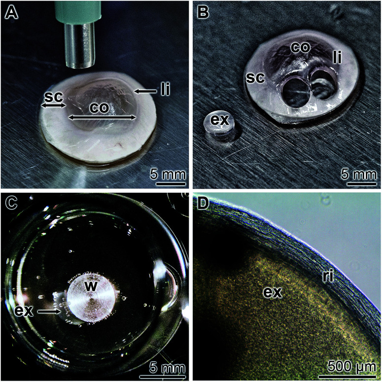
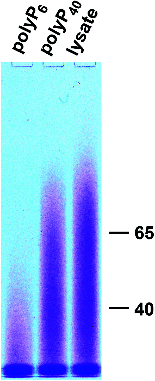
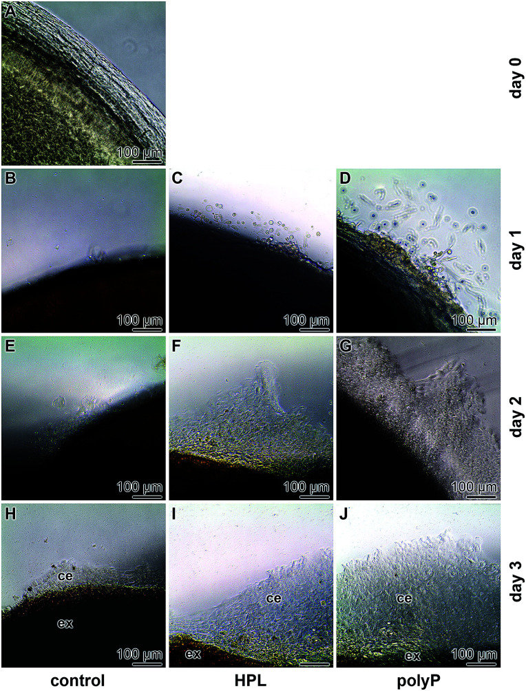
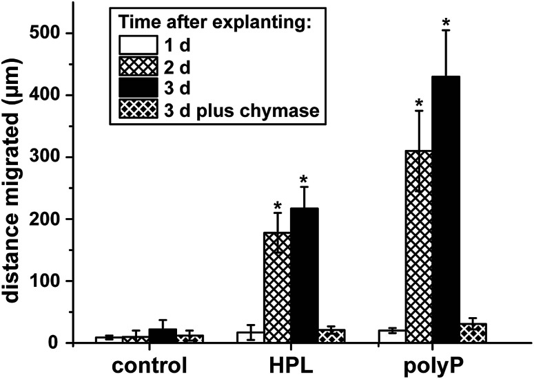
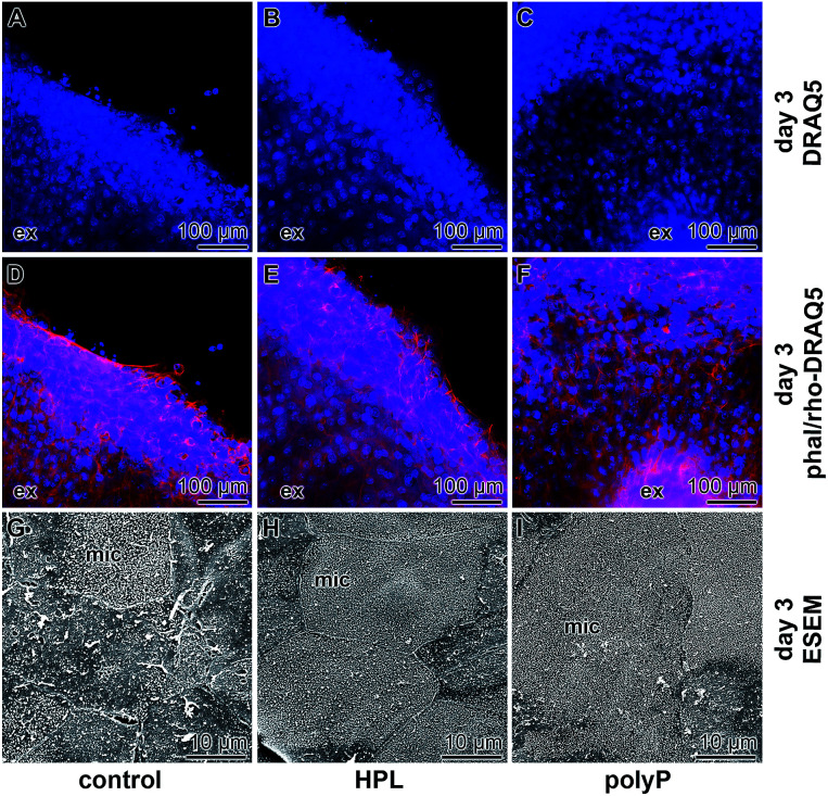
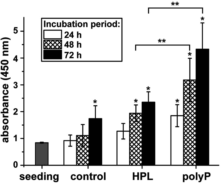
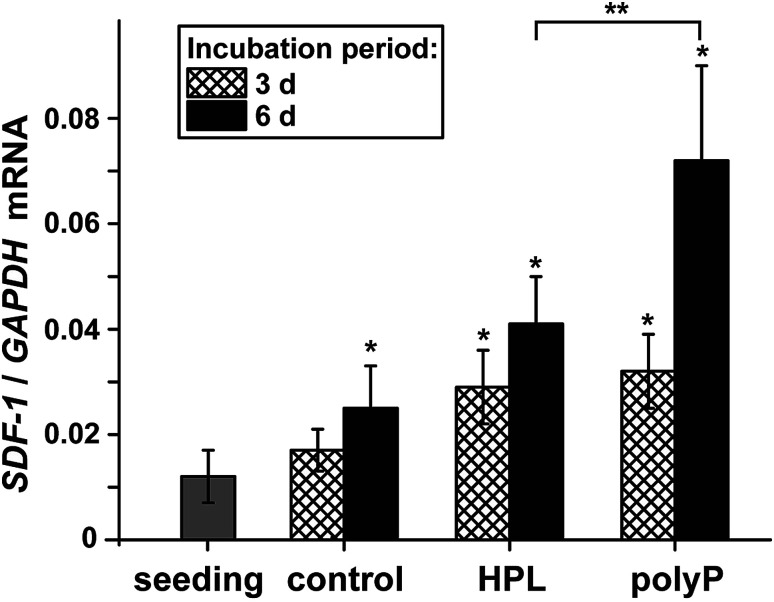
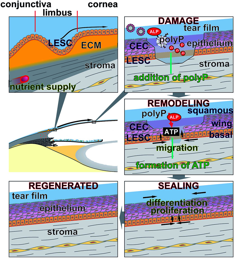
Impaired regeneration of the corneal epithelium, as found in many ocular surface diseases, is a major clinical problem in ophthalmology. We hypothesized that corneal epithelial regeneration can be promoted by the physiological, energy-delivering as well as "morphogenetically active" polymer, inorganic polyphosphate (polyP). Corneal limbal explants (diameter, 4 mm) were cultivated on collagen-coated well plates in the absence or presence of polyP (chain length, ∼40 Pi units; 50 μg ml-1) or human platelet lysate (hp-lysate; 5% v/v). Cell outgrowth and differentiation were analyzed after staining with DRAQ5 (nuclei) and rhodamine phalloidin (cytoskeleton), as well as by environmental scanning electron microscopy (ESEM). Cell growth/viability of hCECs was assessed by XTT assay. The expression of SDF-1 was quantitated by qRT-PCR. Exposure to hp-lysate (also containing polyP) increased cell migration already at day 1. Even stronger was the effect of polyP. This effect was blocked by a mast cell serine protease. The formation of cell multilayers was enhanced by hp-lysate or even more by polyP. ESEM revealed continuous cell junctions and prominent microvilli on the surface of adjacent cells exposed to polyP; those structures were only rarely seen in the controls. The hp-lysate and, more potently, polyP increased the proliferation of hCECs, as well as SDF-1 expression. The findings indicate the potential usefulness of the natural polymer, polyP, for topical treatment of corneal epithelial defects. Future studies are directed to develop suitable formulations of polyP, such as biomimetic polyP nano/microparticles showing an adjustable release kinetics.


Conflict of interest statement

There are no conflicts to declare.

Figures

Fig. 1
Fig. 1. Preparation of human corneal limbal explants. (A and B) Spherical samples from the corneo-scleral rims were withdrawn from with a biopsy punch at the rim between the sclera (sc) and the cornea (co), including also the limbus (li). (C) The explants (ex) obtained were turned around and attached to the bottom of the collagen-coated 24-well plates. The samples were slightly fixed with a stainless steel weight (w). (D) After this procedure no cells were found outside of the rim (ri) of the explants.
Fig. 2
Fig. 2. Identification of polyP in hp-lysate by organic-water extraction and subsequent size separation by urea/polyacrylamide gel electrophoresis; the gels were stained with toluidine blue. The size markers of polyP6 and polyP40 were run in parallel. The most intense staining was found in the hp-lysate polyP at an apparent chain length between 40 and 65 Pi units.
Fig. 3
Fig. 3. Outgrowth of epithelial cells from corneo-scleral explants (ex). (A) At day 0 no cells could be seen in the surrounding of the explants. (B, E and H) Kinetics of outgrowth in the controls, in which no hp-lysate (HPL) or polyP had been added during the 3 d incubation period. In contrast, in the assays which had been supplemented either with (C, F and I) hp-lysate (HPL) or with (D, G and J) polyP an increasing amount of cells (ce) could be visualized.
Fig. 4
Fig. 4. Increase of cells around the explants in control assay or in samples supplemented with hp-lysate (HPL) or polyP. In addition, in the 3 d's experiments the three different explant series were exposed to chymase during the complete incubation period, as described under “Material and methods”. Values are means ± SEM coming from 7 parallel experiments. (*) Significance levels between the migration distances of cells from the HPL or the polyP treated explants and the corresponding controls are marked (P < 0.01).
Fig. 5
Fig. 5. Immunofluorescence and ESEM analysis of the outgrown cells, 3 d after initiating of the assays. In the first series the cell layers outgrown from the explants (ex) were stained with (A–C) DRAQ5 to identify the nuclei or double-stained with (D–F) rhodamine/phalloidin and DRAQ5 to express the complex multilayered arrangement of the cells migrated out from the explants; immunofluorescence light microscopy. The rim of the explant is highlighted (ex). In (G–I) higher magnification images of the epithelial cells showing the different degrees of differentiation of the cells; ESEM. In the controls (G) the cells are not continuously attached to each other with cell-to-cell junctions. In addition, a partial exfoliation of the cells is apparent. In contrast, cells migrated from the explants which had been incubated with hp-lysate (HPL) (H), or with polyP (I) smoothly attach to each other and form a homogeneous margin rim of microvilli on their cell surfaces. Surface areas with extensive microvilli decorations are marked (mic).
Fig. 6
Fig. 6. Metabolic activity/growth of hCECs, growing in ocular epithelia complete medium, during a 72 h incubation period. Cell aliquots were taken after 24 h, 48 h, and 72 h and subjected to the XTT-based proliferation/viability assay. The cultures were treated either with 5% hp-lysate (HPL) or with 50 μg ml−1 Na-polyP/CaCl2 (polyP). Data represent means ± SD of ten independent experiments; the significances between the treated cultures (hp-lysate or polyP) and the respective controls are indicated with one asterisks (*); P < 0.01. The significance values between the correlating treated (hp-lysate and polyP) groups are given with two asterisks (**) (P < 0.01).
Fig. 7
Fig. 7. Steady-state expression levels of the gene encoding for SDF-1 in untreated hCECs, compared to those incubated with 5% hp-lysate (HPL) or with 50 μg ml−1 Na-polyP/CaCl2 (polyP). After incubation for 3 d or 6 d RNA was extracted from the cells and subjected to qRT-PCR analyses. The expression values for SDF-1 were correlated to the expression of GAPDH; the respective expression ratios are given. The results are means from 5 parallel experiments. The (*) significance levels to the expression values in the controls are marked (P < 0.01); the significance correlation between the hp-lysate and polyP are also indicated (**; P < 0.01).
Fig. 8
Fig. 8. Schematic outline of the repair processes proceeding at locally existing defects in the corneal epithelium. The cells which fill the damages originate from stem cells (LESC) in the limbal epithelium; at top left, only the basal layer of the limbus with the LESC without the upper layer of differentiated cells is shown. The LESCs migrate laterally in the impaired epithelium and proliferate and differentiate into the mature corneal epithelial cells (CEC) which form the basal cell-, wing cell-, and the squamous cell layers. These morphogenetic events are augmented by polyP, either existing as Na-polyP or as polyP particles. Mediated by the ALP, which also exists in the tear fluid, extracellular ATP is formed that provides the metabolic energy for the remodeling process. It is proposed that polyP is administered as eye drops.

Similar articles

Cited by

References

    1. Ehlers N. Heegaard S. Hjortdal J. Ivarsen A. Nielsen K. Prause J. U. Acta Ophthalmol. 2010;88:858–861. doi: 10.1111/j.1755-3768.2009.01610.x. - DOI - PubMed
    1. Thoft R. A. Friend J. Invest. Ophthalmol. Visual Sci. 1983;24:1442–1443. - PubMed
    1. Musayeva A. Manicam C. Steege A. Brochhausen C. Straub B. K. Bell K. Pfeiffer N. Gericke A. Am. J. Physiol.: Cell Physiol. 2018;315:C757–C765. doi: 10.1152/ajpcell.00314.2018. - DOI - PubMed
    1. Manicam C. Perumal N. Wasielica-Poslednik J. Ngongkole Y. C. Tschäbunin A. Sievers M. Lisch W. Pfeiffer N. Grus F. H. Gericke A. Sci. Rep. 2018;8:11526. doi: 10.1038/s41598-018-30032-5. - DOI - PMC - PubMed
    1. Haagdorens M. Van Acker S. I. Van Gerwen V. Ní Dhubhghaill S. Koppen C. Tassignon M. J. Zakaria N. Stem Cells Int. 2016;2016:9798374. - PMC - PubMed