

3D Bioprinting for Spinal Cord Injury Repair
- PMID: 35519617
- PMCID: PMC9065470
- DOI: 10.3389/fbioe.2022.847344
3D Bioprinting for Spinal Cord Injury Repair
Abstract
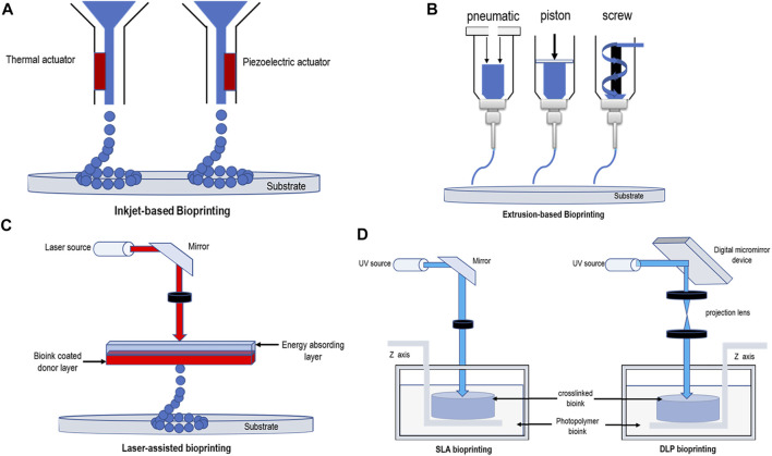
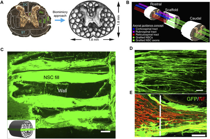
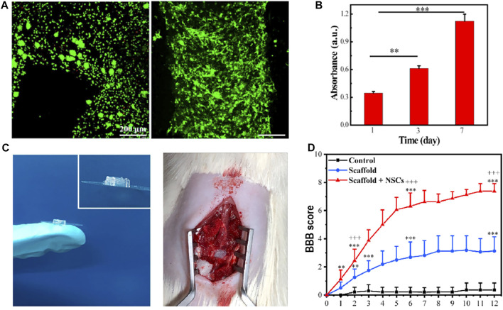
Spinal cord injury (SCI) is considered to be one of the most challenging central nervous system injuries. The poor regeneration of nerve cells and the formation of scar tissue after injury make it difficult to recover the function of the nervous system. With the development of tissue engineering, three-dimensional (3D) bioprinting has attracted extensive attention because it can accurately print complex structures. At the same time, the technology of blending and printing cells and related cytokines has gradually been matured. Using this technology, complex biological scaffolds with accurate cell localization can be manufactured. Therefore, this technology has a certain potential in the repair of the nervous system, especially the spinal cord. So far, this review focuses on the progress of tissue engineering of the spinal cord, landmark 3D bioprinting methods, and landmark 3D bioprinting applications of the spinal cord in recent years.
Keywords: 3D bioprinting; hydrogels; neural system tissue engineering; scaffolds; spinal cord injury repair.
Copyright © 2022 Yuan, Zhang, Yu, Wu and Liu.
Conflict of interest statement
The authors declare that the research was conducted in the absence of any commercial or financial relationships that could be construed as a potential conflict of interest.
Figures
Similar articles
-
The combined application of stem cells and three-dimensional bioprinting scaffolds for the repair of spinal cord injury.Neural Regen Res. 2024 Aug 1;19(8):1751-1758. doi: 10.4103/1673-5374.385842. Epub 2023 Sep 22. Neural Regen Res. 2024. PMID: 38103241 Free PMC article.
-
3D bioprinting applications in neural tissue engineering for spinal cord injury repair.Mater Sci Eng C Mater Biol Appl. 2020 May;110:110741. doi: 10.1016/j.msec.2020.110741. Epub 2020 Feb 19. Mater Sci Eng C Mater Biol Appl. 2020. PMID: 32204049 Review.
-
Development and Application of Three-Dimensional Bioprinting Scaffold in the Repair of Spinal Cord Injury.Tissue Eng Regen Med. 2022 Dec;19(6):1113-1127. doi: 10.1007/s13770-022-00465-1. Epub 2022 Jun 29. Tissue Eng Regen Med. 2022. PMID: 35767151 Free PMC article. Review.
-
3D bioprinted neural tissue constructs for spinal cord injury repair.Biomaterials. 2021 May;272:120771. doi: 10.1016/j.biomaterials.2021.120771. Epub 2021 Mar 25. Biomaterials. 2021. PMID: 33798962
-
Innovative Strategies in 3D Bioprinting for Spinal Cord Injury Repair.Int J Mol Sci. 2024 Sep 4;25(17):9592. doi: 10.3390/ijms25179592. Int J Mol Sci. 2024. PMID: 39273538 Free PMC article. Review.
Cited by
-
Advancements in Antioxidant-Based Therapeutics for Spinal Cord Injury: A Critical Review of Strategies and Combination Approaches.Antioxidants (Basel). 2024 Dec 26;14(1):17. doi: 10.3390/antiox14010017. Antioxidants (Basel). 2024. PMID: 39857350 Free PMC article. Review.
-
Recent trends in spinal trauma management and research.J Clin Orthop Trauma. 2024 Jan 27;49:102351. doi: 10.1016/j.jcot.2024.102351. eCollection 2024 Feb. J Clin Orthop Trauma. 2024. PMID: 38333744 Free PMC article. No abstract available.
-
Current update and trend of 3D printing in spinal surgery: A bibliometric analysis and review of literature.J Orthop. 2023 Nov 29;50:22-28. doi: 10.1016/j.jor.2023.11.054. eCollection 2024 Apr. J Orthop. 2023. PMID: 38162258 Free PMC article. Review.
-
3D printed scaffolds based on hyaluronic acid bioinks for tissue engineering: a review.Biomater Res. 2023 Dec 24;27(1):137. doi: 10.1186/s40824-023-00460-0. Biomater Res. 2023. PMID: 38142273 Free PMC article. Review.
-
The combined application of stem cells and three-dimensional bioprinting scaffolds for the repair of spinal cord injury.Neural Regen Res. 2024 Aug 1;19(8):1751-1758. doi: 10.4103/1673-5374.385842. Epub 2023 Sep 22. Neural Regen Res. 2024. PMID: 38103241 Free PMC article.
References
Publication types
LinkOut - more resources
Full Text Sources