Review
. 2020 Dec 8;10(71):43682-43703.
doi: 10.1039/d0ra08566a. eCollection 2020 Nov 27.

Hydrogels as artificial matrices for cell seeding in microfluidic devices

Affiliations
Review

Hydrogels as artificial matrices for cell seeding in microfluidic devices

Fahima Akther et al. RSC Adv. .

Abstract

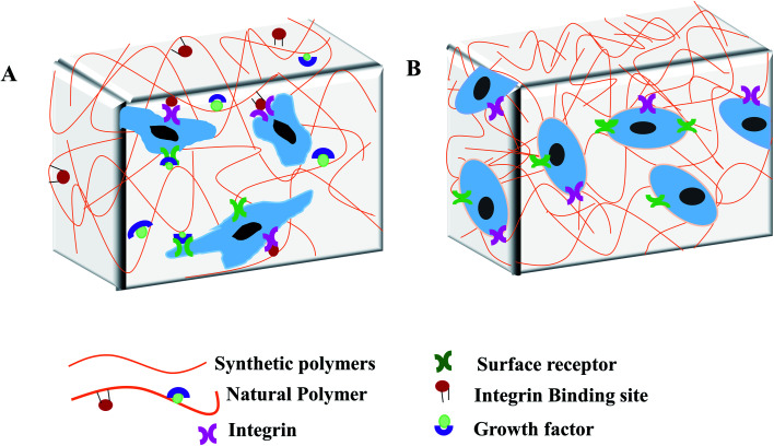
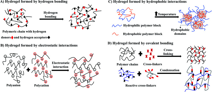
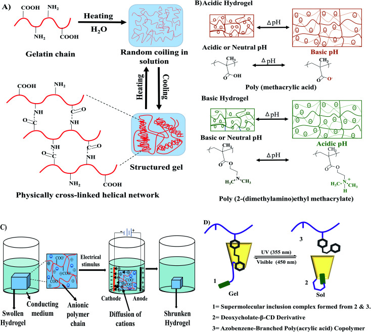
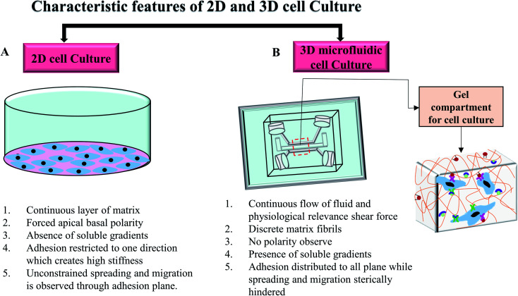
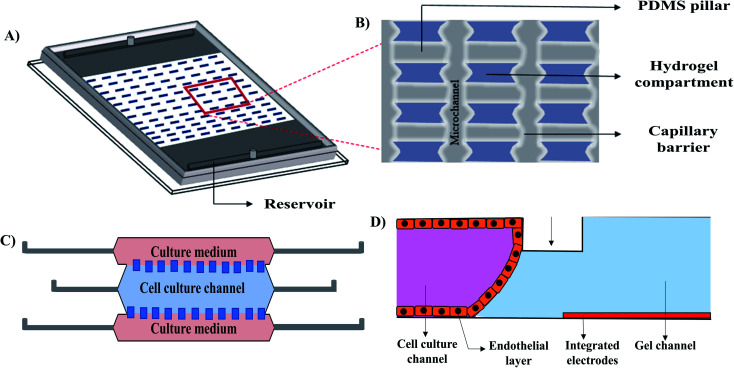
Hydrogel-based artificial scaffolds play a vital role in shifting in vitro models from two-dimensional (2D) cell culture to three-dimensional (3D) cell culture. Microfluidic 3D cell culture systems with a hydrogel matrix encourage biomedical researchers to replace in vivo models with 3D in vitro models with a cellular microenvironment that resembles physiological conditions with greater fidelity. Hydrogels can be designed as an artificial extracellular matrix scaffold for providing spatial orientation and promoting cellular interactions with surroundings. Selecting the appropriate hydrogels and their fabrication techniques are the key to mimic the in vivo mechanical environment. Moreover, combining a microfluidic technique with a hydrogel-based 3D cell culture system can create a complex and controlled microenvironment for the cells by placing small biosamples inside the microchannel. This paper provides an overview of the structural similarities of the hydrogels as an extracellular matrix (ECM), their classification and fabrication techniques as an ECM, and their use in microfluidic 3D cell culture systems. Finally, the paper presents the current challenges and future perspectives of using hydrogel scaffolds in microfluidic 3D cell culture systems.


Conflict of interest statement

Nothing to declare.

Figures

Fig. 1
Fig. 1. Structural similarities between extracellular matrix (ECM) and hydrogels. (A) ECM is consisted of three groups of macromolecules: structural proteins (collagens), proteoglycans, and glycoproteins (fibronectin) to form the matrix for cell attachment. Proteoglycan and structural fibrous proteins fill the cells' interstitial space to provide the mechanical and biological supports. Hydrogel scaffold from (B) natural polymers and (C) synthetic polymers. Fibrous hydrogel polymers link by interconnected microscopic pores to provide mechanical and biological cellular support.
Fig. 2
Fig. 2. Hydrogel based artificial scaffold for cell culture. (A) Scaffold, composed of natural polymers, provides the cellular support by enabling the cells to bind with a variety of different integrin-binding sites and growth factor and regulates cell behaviour through activating signalling cascade. (B) Scaffold, composed of synthetic polymers, lacks growth factors and integrin binding sites that only provides the mechanical support for cell growth.
Fig. 3
Fig. 3. Schematic illustration of synthesis of hydrogel by physical methods (A–C) and chemical method (D).
Fig. 4
Fig. 4. Stimuli-responsive hydrogels. (A) Thermoresponsive sol–gel transition of gelatin. (B) pH-sensitive swelling–deswelling behaviour of acidic and basic hydrogels redrew from Kocak et al. (D) Influence of applied electric filed on hydrogel reconstructed from Qureshi et al. (C) Photosensitivity in the sol–gel transition of azobenzene-based hydrogel redrew from Zhao et al.
Fig. 5
Fig. 5. Characteristic attributes of 2D and 3D microfluidic cell culture systems. (A) Characteristics of the conventional 2D cell culture system. (B) A commonly used microfluidic 3D cell culture model and cellular orientation in the gel compartment.
Fig. 6
Fig. 6. Reconstructed schematic diagrams of 3D microfluidic cell culture devices. (A) Isometric view of the hydrogel patterns with a zoomed in illustration (B) of the capillary barriers, PDMS pillars and hydrogel compartments of the compartmentalized model, (C) TEM (the tumour microenvironment) model, and (D) vascular model.

Similar articles

Cited by

References

    1. Rozario T. Desimone D. W. The extracellular matrix in development and morphogenesis: a dynamic view. Dev. Biol. 2010;341(1):126–140. doi: 10.1016/j.ydbio.2009.10.026. - DOI - PMC - PubMed
    1. Steven R. C. Jason A. B. A practical guide to hydrogels for cell culture. Nat. Methods. 2016;13(5):405. doi: 10.1038/nmeth.3839. - DOI - PMC - PubMed
    1. Kapałczyńska M. Kolenda T. Przybyła W. Zajączkowska M. Teresiak A. Filas V. et al., 2D and 3D cell cultures - a comparison of different types of cancer cell cultures. Arch. Med. Sci. 2018;14(4):910–919. - PMC - PubMed
    1. Lv D. Yu S.-C. Ping Y.-F. Wu H. Zhao X. Zhang H. et al., A three-dimensional collagen scaffold cell culture system for screening anti-glioma therapeutics. Oncotarget. 2016;7(35):56904–56914. doi: 10.18632/oncotarget.10885. - DOI - PMC - PubMed
    1. Lutolf M. P. Hubbell J. A. Synthetic biomaterials as instructive extracellular microenvironments for morphogenesis in tissue engineering. Nat. Biotechnol. 2005;23:47–55. doi: 10.1038/nbt1055. - DOI - PubMed