Retracted Article: Sauchinone inhibits high glucose-induced oxidative stress and apoptosis in retinal pigment epithelial cells
- PMID: 35519842
- PMCID: PMC9064550
- DOI: 10.1039/c9ra02817j
Retracted Article: Sauchinone inhibits high glucose-induced oxidative stress and apoptosis in retinal pigment epithelial cells
Retraction in
-
Retraction: Sauchinone inhibits high glucose-induced oxidative stress and apoptosis in retinal pigment epithelial cells.RSC Adv. 2021 Jan 28;11(9):5243. doi: 10.1039/d1ra90044g. eCollection 2021 Jan 25. RSC Adv. 2021. PMID: 35427013 Free PMC article.
Abstract
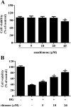
Diabetic retinopathy (DR) is a common complication of diabetes mellitus and results in acquired blindness among working-age adults. It has been demonstrated that high glucose (HG)-induced oxidative stress and cell apoptosis in retinal pigment epithelial (RPE) cells are major factors for the pathogenesis of DR. Sauchinone, one of the active lignan isolated from Saururus chinensis, was reported to possess anti-oxidant and anti-apoptosis effects. In the present study, we investigated the effects of sauchinone on HG-induced oxidative stress and apoptosis in ARPE-19 cells. Our results proved that sauchinone improved the cell viability of HG-induced ARPE-19 cells. Moreover, sauchinone treatment caused a decrease in intracellular reactive oxygen species (ROS) generation and an increase in the activities of superoxide dismutase (SOD), glutathione peroxidase (GPx), and catalase (CAT). Besides, flow cytometry showed that the apoptotic rate in sauchinone-treated ARPE-19 cells obviously decreased as compared in the HG-treated cells. Western blot indicated that sauchinone treatment caused a significant decrease in bax expression and increase in bcl-2 expression in HG-treated ARPE-19 cells. Sauchinone treatment enhanced the HG-caused induction of p-Akt, nuclear factor erythroid 2-related factor (Nrf2), and heme oxygenase-1 (HO-1) expressions in ARPE-19 cells. However, the inhibitor of Akt, LY294002, reversed the effects of sauchinone on cell viability, oxidative stress, and cell apoptosis in HG-treated ARPE-19 cells. These findings suggested that sauchinone treatment prevented HG-induced oxidative stress and apoptosis via regulating the Akt/Nrf2/HO-1 pathway in HG-induced RPE cells. These findings suggested that sauchinone might be a therapeutic agent for the treatment and prevention of DR.
This journal is © The Royal Society of Chemistry.
Conflict of interest statement
The authors declare that there are no conflicts of interest.
Figures


Similar articles
-
Polygonatum sibiricum polysaccharide inhibits high glucose-induced oxidative stress, inflammatory response, and apoptosis in RPE cells.J Recept Signal Transduct Res. 2022 Apr;42(2):189-196. doi: 10.1080/10799893.2021.1883061. Epub 2021 Feb 8. J Recept Signal Transduct Res. 2022. PMID: 33554697
-
Knockdown of ALK7 inhibits high glucose-induced oxidative stress and apoptosis in retinal pigment epithelial cells.Clin Exp Pharmacol Physiol. 2020 Feb;47(2):313-321. doi: 10.1111/1440-1681.13189. Epub 2019 Nov 19. Clin Exp Pharmacol Physiol. 2020. PMID: 31608496
-
CTRP3 inhibits high glucose-induced oxidative stress and apoptosis in retinal pigment epithelial cells.Artif Cells Nanomed Biotechnol. 2019 Dec;47(1):3758-3764. doi: 10.1080/21691401.2019.1666864. Artif Cells Nanomed Biotechnol. 2019. PMID: 31556307
-
Aloperine protects human retinal pigment epithelial cells against hydrogen peroxide-induced oxidative stress and apoptosis through activation of Nrf2/HO-1 pathway.J Recept Signal Transduct Res. 2022 Feb;42(1):88-94. doi: 10.1080/10799893.2020.1850787. Epub 2020 Nov 30. J Recept Signal Transduct Res. 2022. PMID: 33256538
-
Benzoxanthenone Lignans Related to Carpanone, Polemanone, and Sauchinone: Natural Origin, Chemical Syntheses, and Pharmacological Properties.Molecules. 2025 Apr 10;30(8):1696. doi: 10.3390/molecules30081696. Molecules. 2025. PMID: 40333626 Free PMC article. Review.
Cited by
-
Mechanistic insights into the alterations and regulation of the AKT signaling pathway in diabetic retinopathy.Cell Death Discov. 2023 Nov 17;9(1):418. doi: 10.1038/s41420-023-01717-2. Cell Death Discov. 2023. PMID: 37978169 Free PMC article. Review.
-
Nature against Diabetic Retinopathy: A Review on Antiangiogenic, Antioxidant, and Anti-Inflammatory Phytochemicals.Evid Based Complement Alternat Med. 2022 Mar 9;2022:4708527. doi: 10.1155/2022/4708527. eCollection 2022. Evid Based Complement Alternat Med. 2022. PMID: 35310030 Free PMC article. Review.
References
Publication types
LinkOut - more resources
Full Text Sources
Research Materials
Miscellaneous

