. 2019 Jan 31;9(8):4172-4179.
doi: 10.1039/c8ra10390a. eCollection 2019 Jan 30.

Enhancement of protein stability by an additional disulfide bond designed in human neuroglobin

Affiliations

Enhancement of protein stability by an additional disulfide bond designed in human neuroglobin

Hai-Xiao Liu et al. RSC Adv. .

Abstract

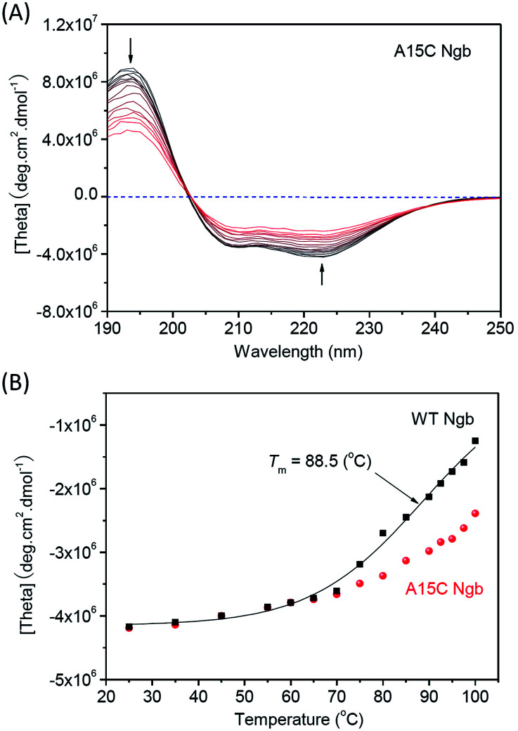
Human neuroglobin (Ngb) forms an intramolecular disulfide bond between Cys46 and Cys55, with a third Cys120 near the protein surface, which is a promising protein model for heme protein design. In order to protect the free Cys120 and to enhance the protein stability, we herein developed a strategy by designing an additional disulfide bond between Cys120 and Cys15 via A15C mutation. The design was supported by molecular modeling, and the formation of Cys15-Cys120 disulfide bond was confirmed experimentally by ESI-MS analysis. Molecular modeling, UV-Vis and CD spectroscopy showed that the additional disulfide bond caused minimal structural alterations of Ngb. Meanwhile, the disulfide bond of Cys15-Cys120 was found to enhance both Gdn·HCl-induced unfolding stability (increased by ∼0.64 M) and pH-induced unfolding stability (decreased by ∼0.69 pH unit), as compared to those of WT Ngb with a single native disulfide bond of Cys46-Cys55. Moreover, the half denaturation temperature (T m) of A15C Ngb was determined to be higher than 100 °C. In addition, the disulfide bond of Cys15-Cys120 has slight effects on protein function, such as an increase in the rate of O2 release by ∼1.4-fold. This study not only suggests a crucial role of the artificial disulfide in protein stabilization, but also lays the groundwork for further investigation of the structure and function of Ngb, as well as for the design of other functional heme proteins, based on the scaffold of A15C Ngb with an enhanced stability.


Conflict of interest statement

There are no conflicts to declare.

Figures

Fig. 1
Fig. 1. (A) Modeling structure of A15C Ngb, showing the disulfide bonds (indicated by circle lines), and (B) overlaid with the X-ray structure of WT Ngb (blue), with the secondary structures of HA–HH labeled.
Fig. 2
Fig. 2. Spectroscopic properties of A15C Ngb. (A) UV-Vis spectra in both oxidation states, and (B) CD spectrum in ferric state (10 μM protein in 100 mM potassium phosphate buffer, pH 7.0, 25 °C), with WT Ngb shown for comparison.
Fig. 3
Fig. 3. ESI-MS analysis of WT Ngb (A) and A15C Ngb (B) as purified (left) and treated with reducing agent TCEP (right).
Fig. 4
Fig. 4. UV-Vis spectra of Gdn·HCl-induced unfolding of (A) A15C Ngb (10 μM protein in 100 mM potassium phosphate buffer, pH 7.0). (B) Normalized absorbance of the Soret band versus Gdn·HCl concentrations.
Fig. 5
Fig. 5. UV-Vis spectra of acid-induced unfolding of (A) A15C Ngb (10 μM). (B) Normalized absorbance of the Soret band versus pH values.
Fig. 6
Fig. 6. CD spectra of thermal-induced unfolding of (A) A15C Ngb (10 μM protein in 100 mM potassium phosphate buffer, pH 7.0). (B) Plots of absorbance at 222 nm versus temperature.
Fig. 7
Fig. 7. UV-Vis spectra of thermal-induced unfolding of (A) WT Ngb and (B) A15C Ngb (10 μM protein in 100 mM potassium phosphate buffer, pH 7.0). The plot of Soret band at 413 nm versus temperature was shown as an inset.
Fig. 8
Fig. 8. (A) UV-Vis spectra of autoxidation of oxy-A15C Ngb (10 μM protein in 100 mM potassium phosphate buffer, pH 7.0) for 60 min at 25 °C. (B) Stopped-flow spectra of oxy-A15C Ngb (5 μM protein in 100 mM potassium phosphate buffer, pH 7.0), in reaction with 1 mM dithionite in the same buffer for 10 s at 25 °C. The time dependent changes for the oxy-form at 542 nm were shown as insets, with WT Ngb under the same conditions shown for comparison.

Similar articles

Cited by

References

    1. Lu Y. Yeung N. Sieracki N. Marshall N. M. Nature. 2009;460:855–862. doi: 10.1038/nature08304. - DOI - PMC - PubMed
    1. Du J. Sono M. Dawson J. H. Coord. Chem. Rev. 2011;255:700–716. doi: 10.1016/j.ccr.2011.01.029. - DOI - PMC - PubMed
    1. Liebl U. Lambry J.-C. Vos M. H. Biochim. Biophys. Acta. 2013;1834:1684–1692. doi: 10.1016/j.bbapap.2013.02.025. - DOI - PubMed
    1. Poulos T. L. Chem. Rev. 2014;114:3919–3962. doi: 10.1021/cr400415k. - DOI - PMC - PubMed
    1. Liu J. Chakraborty S. Hosseinzadeh P. Yu Y. Tian S. Petrik I. Bhagi A. Lu Y. Chem. Rev. 2014;114:4366–4469. doi: 10.1021/cr400479b. - DOI - PMC - PubMed