. 2022 Feb 14;4(5):342-361.
doi: 10.1096/fba.2021-00055. eCollection 2022 May.

RhoA-ROCK competes with YAP to regulate amoeboid breast cancer cell migration in response to lymphatic-like flow

Affiliations

RhoA-ROCK competes with YAP to regulate amoeboid breast cancer cell migration in response to lymphatic-like flow

Amina Mohammadalipour et al. FASEB Bioadv. .

Abstract

Lymphatic drainage generates force that induces prostate cancer cell motility via activation of Yes-associated protein (YAP), but whether this response to fluid force is conserved across cancer types is unclear. Here, we show that shear stress corresponding to fluid flow in the initial lymphatics modifies taxis in breast cancer, whereas some cell lines use rapid amoeboid migration behavior in response to fluid flow, a separate subset decrease movement. Positive responders displayed transcriptional profiles characteristic of an amoeboid cell state, which is typical of cells advancing at the edges of neoplastic tumors. Regulation of the HIPPO tumor suppressor pathway and YAP activity also differed between breast subsets and prostate cancer. Although subcellular localization of YAP to the nucleus positively correlated with overall velocity of locomotion, YAP gain- and loss-of-function demonstrates that YAP inhibits breast cancer motility but is outcompeted by other pro-taxis mediators in the context of flow. Specifically, we show that RhoA dictates response to flow. GTPase activity of RhoA, but not Rac1 or Cdc42 Rho family GTPases, is elevated in cells that positively respond to flow and is unchanged in cells that decelerate under flow. Disruption of RhoA or the RhoA effector, Rho-associated kinase (ROCK), blocked shear stress-induced motility. Collectively, these findings identify biomechanical force as a regulator amoeboid cell migration and demonstrate stratification of breast cancer subsets by flow-sensing mechanotransduction pathways.

Keywords: ECM–receptor interactions; actomyosin cytoskeleton; biomechanical force; hippo; mechanotransduction; motility; shear stress.


Conflict of interest statement

The authors have declared explicitly that there are no conflicts of interest in connection with this article.

Figures

FIGURE 1
FIGURE 1
Flow can promote or inhibit motility in breast cancer cell lines. (A) Tracking analysis shows that cellular velocity is altered by flow in various breast cancer cell lines. Wall shear stress (WSS) exposure significantly increases motility in EFM‐19, MDA‐MB‐231, and MDA‐MB‐415 cells; whereas, WSS inhibits migration in MCF‐7 and HCC1187 lines. HCC1806 exhibits no detectable change in motility. Results are expressed as mean ± SD; n ≥ 70 individual cells for all lines (unpaired two‐tailed t‐test, ***p < 0.001). (B, C) Still photos from time‐lapse movie files and representative cumulative plots show that cell migration radiates from the origin of each cell's starting location with no overt directionality. (D) Magnified view of single MDA‐MB‐231 cell undergoing a shift toward amoeboid morphology within minutes of initiation of WSS. Purple pseudocolor encompasses cell body and membrane projections. Yellow dot and lines mark starting location and path of locomotion
FIGURE 2
FIGURE 2
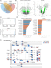
Positive and negative motility response defines transcriptionally distinct subsets of cancer cells. (A) Venn diagram illustrates overlap and uniqueness of gene expression signatures induced by lymphatic intensities of wall shear stress (WSS) in PC3 prostate cancer cells and two subsets of breast cancer cells defined by increased (positive) or decreased (negative) migration response under flow. The positive versus negative comparison contrasts the two breast cancer subsets. (B) Differentially expressed genes are depicted by volcano plot with cutoffs demarcated by dashed lines and color at p‐value < 0.05 and −1 < log2 fold change < 1. (C) Transcripts of genes representative of those regulated in distinct directions in the breast cancer subsets. (D) GSEA of top 20 KEGG pathways show broad changes in adhesion, cytokine signaling, and cytoskeleton. Contrast reflects change relative to static conditions such that orange bars indicate pathways upregulated in positive breast cancer responders exposed to flow relative to negative responders exposed to flow. Blue bars depict pathways predicted to be more highly downregulated in positive responders relative to negative responders. Prostate cancer pathways reflect WSS versus static conditions. (E) Expression of HIPPO pathway components are shown to indicate fold change in positive versus negative breast cancer comparison in the left half of each split node and prostate on the right of each node. Color scale legends indicate fold change in gene expression following 6 h of WSS in breast or prostate
FIGURE 3
FIGURE 3
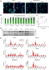
YAP nuclear localization positively correlates with motility. (A) Representative images of immunofluorescent staining show altered YAP subcellular localization upon exposure to flow. Scale bar represents 25 μm. (B) Quantification demonstrates that the fraction of cells containing YAP in the nucleus decreases in MCF‐7 but increases in other lines, such as MDA‐MB‐231 and MDA‐MB‐415. Grey background demarcates cancer lines with increased frequency of cells containing nuclear YAP under flow (N > C and N = C). Error bars represent SEM. (C) Fold change in distance traveled and nuclear YAP are significantly correlated across all cell lines (Linear regression, p = 0.04). (D) Dephosphorylation of YAP S127 is triggered by flow in MCF‐7, EFM‐19, MDA‐MB‐231, and MDA‐MB‐415 cell lines. (E) Transcript levels of seven genes classically used as readouts of YAP and/or TAZ activity, such as CTGF, were measured at 1, 6, or 12 h after initiation of wall shear stress. Expression of these genes was stimulated by flow to varying extents in all breast lines tested (one‐way ANOVA, *p < 0.05 relative to static control). Metalloprotease genes MMP2 and MMP9 were also evaluated as indicators of invasive potential. Error bars represent SEM
FIGURE 4
FIGURE 4
YAP constitutive activation inhibits whereas knockdown promotes breast cancer cell migration. (A) Ectopic expression of constitutively active forms of YAP and TAZ by transient transfection of plasmids used in our prior study of prostate cancer failed to induce motility under static conditions. Instead, constitutively active YAP S127A suppressed taxis (one‐way ANOVA Dunn's, *p < 0.05). pEGFP‐N1 served as the control vector. (B) As a complementary approach, retrovirus was used to express YAP S127A,S381A in MDA‐MB‐231. Blot shows that FLAG tag is detectable even when virus is diluted 1:20 during infection. The MSCV‐IRES‐Hygro control vector is demarcated as IH. (C, D) Introduction of constitutively active forms of YAP by retrovirus, including S127A and S127A,S381A mutants, were confirmed to result in elevated total YAP protein levels and suppressed migration speed in MDA‐MB‐231 cells (one‐way ANOVA, **p < 0.01 relative to uninfected control, #p < 0.05 relative to IH control). (E, F) Expression of constitutively active YAP was also successful in MCF‐7 cells and similarly reduced migration as opposed to increasing movement (one‐way ANOVA, **p < 0.01 relative to uninfected control, #p < 0.05 relative to IH empty vector control). (G, H) Knockdown of YAP by three independent shRNAs did not profoundly alter motility response to WSS by MDA‐MB‐231. Instead, YAP depletion increased cellular velocity under static conditions, along with a proportionate increase under WSS (two‐way ANOVA, *p < 0.05, **p < 0.01). MSCV‐Zsgreen‐2A‐Puro‐shRNA‐FF served as the control for retroviral knockdown. (I, J) MCF‐7 also continued to respond to WSS with reduced motility with or without YAP knockdown (two‐way ANOVA, *p < 0.05, **p < 0.01). (K) siRNAs were used as independent validation to knockdown YAP and TAZ singly and in combination. (L) siRNA‐based YAP or TAZ knockdown alone did not produce amplified migration speeds, but YAP‐TAZ combination knockdown increased motility (two‐way ANOVA, *p < 0.05). Error bars on motility plots represent SD
FIGURE 5
FIGURE 5
RhoA‐ROCK activity determines motility response to flow. (A) Focal adhesion was identified as a top KEGG pathway differentially regulated between positive and negative breast cancer subsets. Fold change in pathway signaling effectors is depicted for positive versus negative breast cancer contrast in the left half of split nodes and for prostate on the right of the node. Color scale legends indicate fold change after 6 h of wall shear stress (WSS). (B) Flow significantly elevated GTP‐bound (active) RhoA levels 60 min after initiation of WSS in MDA‐MB‐231 (unpaired t‐test, *p < 0.05). RhoA GTPase activity was unchanged in MCF‐7. Error bars represent SEM. (C) Rac1 activity was not significantly changed in either MDA‐MB‐231 or MCF‐7. (D) Cdc42 activity was unchanged in response to flow in both cell lines. (E, F) WSS‐induced motility in MDA‐MB‐231 is suppressed by ROCK inhibitor Y27632 (10 µm) and disruption of RhoA activity by dominant negative or constitutively active forms of RhoA (two‐way ANOVA, *p < 0.05). Error bars represent SD

References

    1. Biswas AK, Acharyya S. Understanding cachexia in the context of metastatic progression. Nat Rev Cancer. 2020;20(5):274‐284. 10.1038/s41568-020-0251-4 - DOI - PubMed
    1. Balkwill FR, Capasso M, Hagemann T. The tumor microenvironment at a glance. J Cell Sci. 2012;125(23):5591‐5596. doi:10.1242/jcs.116392 - DOI - PubMed
    1. Jain RK. Normalizing tumor microenvironment to treat cancer: bench to bedside to biomarkers. J Clin Oncol. 2013;31(17):2205‐2218. doi:10.1200/JCO.2012.46.3653 - DOI - PMC - PubMed
    1. Jaalouk DE, Lammerding J. Mechanotransduction gone awry. Nat Rev Mol Cell Biol. 2009;10(1):63‐73. doi:10.1038/nrm2597 - DOI - PMC - PubMed
    1. Paduch R. The role of lymphangiogenesis and angiogenesis in tumor metastasis. Cell Oncol. 2016;39(5):397‐410. doi:10.1007/s13402-016-0281-9 - DOI - PMC - PubMed