. 2020 May 27;10(34):20118-20128.
doi: 10.1039/d0ra04090h. eCollection 2020 May 26.

Mussel inspired ZIF8 microcarriers: a new approach for large-scale production of stem cells

Affiliations

Mussel inspired ZIF8 microcarriers: a new approach for large-scale production of stem cells

Mahsa Asadniaye Fardjahromi et al. RSC Adv. .

Abstract

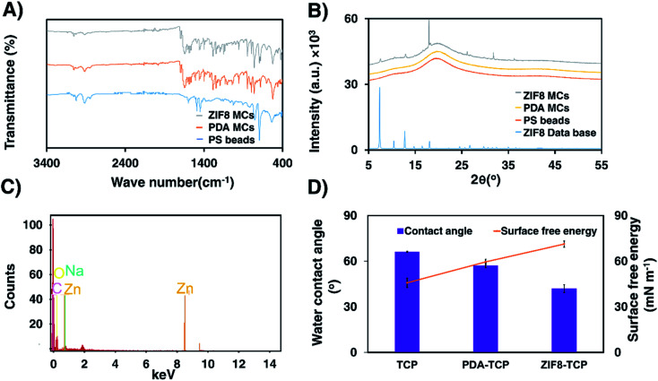
Metal-organic frameworks (MOFs) have high porosity, large surface area, and tunable functionality and have been widely used for drug loading. The aim of this study was to exploit unique features of zeolitic imidazolate framework-8 (ZIF8) to develop an innovative composite microcarrier (MC) for human mesenchymal stem cells (hMSCs) adhesion and proliferation. ZIF8 MCs were prepared by immobilization of polydopamine/polyethyleneimine (PDA/PEI) and ZIF8 on the surface of polystyrene beads. The chemical properties of MCs such as coating stability and homogeneity were characterized by different techniques such as ATR-FTIR, XRD, EDX, SEM, and contact angle. The prepared MCs were tested using human adipose-derived mesenchymal stem cells (hADSCs) in both static and dynamic conditions, and results were compared to a commercially available MC (Star-Plus), polydopamine coated MCs and amine-functionalized MCs as a control. Results show that PDA/PEI/ZIF8 coated MCs (in short: ZIF8 MCs) provides an excellent biocompatible environment for hADSCs adhesion and growth. In conclusion, ZIF8 MCs represent suitable and low-cost support for hADSCs culture and expansion, which can maximize cell yield and viability while preserving hADSCs multipotency. The present findings have revealed this strategy has the potential for chemical and topographical modification of MCs in tissue engineering applications.


Conflict of interest statement

There are no conflicts to declare.

Figures

Scheme 1
Scheme 1. Schematic fabrication of bioinspired MCs and expansion of hADSCs in static and dynamic conditions. PDA MCs were synthesized by immersing PS beads on Tris base buffer solution in the presence of PEI and DA·HCl. ZIF8 MCs, then fabricated by 3D coating ZIF8 thin film on the surface of PDA MCs in a solution containing (2-MIM) and Zn(NO3)2. Cell culture was studied with two methods: static two-dimensional (2D), and three-dimensional dynamic (3DD). In the static method, hADSCs were seeded on MCs in 24-well ultra-low attachment plate and cultured for 7 days. On the dynamic condition, cell culture has been performed in PBS bioreactor and the suspension of hADSCs and MCs were stirred for 7 days.
Fig. 1
Fig. 1. Light microscopy images of (A) PS beads, (B) PDA MCs, (C) ZIF8 MCs. Digital images of each sample are presented as inset color of PS beads changed to dark brown after coating with PDA. 3D AFM topographical images of (D)TCP, (E) PDA-TCP, (F) ZIF8-TCP. Surface roughness changed after coating PDA/PEI and PDA/PEI/ZIF8 on the surface of TCP.
Fig. 2
Fig. 2. Characterization of PDA MCs and ZIF8 MCs. (A) ATR-FTIR spectra of PS beads, PDA MCs and ZIF8 MCs. (B) XRD patterns of PS beads, PDA MCs, ZIF8 MCs, and ZIF8 database. (C) Energy dispersive spectra (EDX) of ZIF8 MCs. (D) Water contact angle and surface free energy measurement of TCP, PDA-TCP and ZIF8-TCP with diiodomethane, water and glycerol using Van Oss method. Each sample was assessed in three replicates.
Fig. 3
Fig. 3. SEM images of PS beads (A1–A3), PDA MCs (B1–B3), ZIF8 MCs (C1–C3). Surface roughness increased by coating ZIF8/PDA/PEI on the surface of PS beads.
Fig. 4
Fig. 4. Fluorescent microscopy images of hADSCs attachment and growth on MCs under static condition over 7 days. PS beads: (A1) day 1, (A2) day 3, (A3) day 7. PDA MC: (B1) day 1, (B2) day 3, (B3) day 7. ZIF8 MC: (C1) day 1, (C2) day 3, (C3) day 7. Star-Plus MC: (D1) day 1, (D2) day 3, (D3) day 7. Cell adhesion increased by coating PDA and PDA/ZIF8 on the surface of PS beads. ZIF8 MC enabled to support and proliferate cells with high efficiency. The nucleus was stained with DAPI. Scale bar: 100 μm.
Fig. 5
Fig. 5. Confocal microscopy images of MCs in dynamic conditions. PS beads: (A1) day 1, (A2) day 3, (A3) day 7. PDA MC: (B1) day 1, (B2) day 3, (B3) day 7. ZIF8 MC: (C1) day 1, (C2) day 3, (C3) day 7. Star-Plus MCs: (D1) day 1, (D2) day 3, (D3) day 7. The images of PS beads and PDA MCs in different days of culture indicate cell proliferation enhanced by PDA coating. Using ZIF8 thin film layer in the fabrication of MCs show a significant increase in cell attachment and growth compared to PS beads and PDA MCs. The nuclei were stained with DAPI. Yellow scale bars indicate 100 μm, and white scale bars indicate 50 μm.
Fig. 6
Fig. 6. Potential of proliferation and differentiation of hMSCs cultured in MCs in 3D cell culture. (A) Total viable cell density obtained by 3D expansion of cells on PS MCs, PDA MCs, ZIF8 MCs, and Star-Plus MCs after 7 days of culture. ZIF8 MCs supported a high level of ADSCs (5.8 fold expansion). (B) Analysis of cell attachment and growth on the surface of PS beads, PDA MCs, ZIF8 MCs, and Star-Plus MCs using MTS assay. PDA and ZIF8 coating improve cell attachment and growth of PS beads with a significant difference. Multipotency assay of harvested hADSCs from (C) PDA MCs (osteoblasts, left panel) (adipocytes, right panel) and (D) (osteoblasts, left panel) (adipocytes, right panel). PDA and ZIF8 MCs retained their differential potential under dynamic conditions. Data represented as mean ± SEM, from four independent experiments, numerical variables were statistically evaluated by one-way ANOVA test.*P value < 0.05 considered significant. The result for the above indicates a high significant **P value < 0.01, ***P value < 0.001, ****P value < 0.0001.

References

    1. Fitzsimmons R. E. Mazurek M. S. Soos A. Simmons C. A. Stem Cells Int. 2018;2018:8031718. doi: 10.1155/2018/8031718. - DOI - PMC - PubMed
    1. Jiang Y. Jahagirdar B. N. Reinhardt R. L. Schwartz R. E. Keene C. D. Ortiz-Gonzalez X. R. Reyes M. Lenvik T. Lund T. Blackstad M. Nature. 2002;418:41–49. doi: 10.1038/nature00870. - DOI - PubMed
    1. Sekula M. and Zuba-Surma E. K., Biomaterials in Regenerative Medicine, 2018, pp. 361–375
    1. Vinatier C. Guicheux J. Ann. Phys. Rehabil. Med. 2016;59:139–144. doi: 10.1016/j.rehab.2016.03.002. - DOI - PubMed
    1. Rizvanov A. A. Persson J. Şahin F. Bellusci S. Oliveira P. J. Stem Cells Int. 2016;2016:3157365. - PMC - PubMed