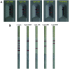
Ultrasensitive strips for the quadruple detection of nitrofuran metabolite residues
- PMID: 35520510
- PMCID: PMC9059960
- DOI: 10.1039/c8ra10589h
Ultrasensitive strips for the quadruple detection of nitrofuran metabolite residues
Abstract
In this biosensor system, metabolite residues were derived by using a previous B-CBA synthesis method to label a biotin moiety for enrichment by streptavidin coated magnetic beads. Antibodies specific for derivatives were conjugated with carboxyl-modified barcode DNAs which were used as templates for strand displacement amplification (SDA). The assay can detect trace levels of 7.20 ppt of SEM, 11.58 ppt of AHD, 7.24 ppt of AOZ and 2.31 ppt of AMOZ, respectively.
This journal is © The Royal Society of Chemistry.
Conflict of interest statement
There are no conflicts to declare.
Figures


Similar articles
-
Lateral flow biosensor for multiplex detection of nitrofuran metabolites based on functionalized magnetic beads.Anal Bioanal Chem. 2016 Sep;408(24):6703-9. doi: 10.1007/s00216-016-9787-2. Epub 2016 Jul 20. Anal Bioanal Chem. 2016. PMID: 27438720
-
Freeze-synthesized bio-barcode immunoprobe based multiplex fluorescence immunosensor for simultaneous determination of four nitrofuran metabolites.Food Chem. 2022 Nov 1;393:133424. doi: 10.1016/j.foodchem.2022.133424. Epub 2022 Jun 7. Food Chem. 2022. PMID: 35751212
-
A multiplex immunochromatographic test using gold nanoparticles for the rapid and simultaneous detection of four nitrofuran metabolites in fish samples.Anal Bioanal Chem. 2018 Jan;410(1):223-233. doi: 10.1007/s00216-017-0714-y. Epub 2017 Oct 30. Anal Bioanal Chem. 2018. PMID: 29085985
-
Quantum Dot Nanobeads as Multicolor Labels for Simultaneous Multiplex Immunochromatographic Detection of Four Nitrofuran Metabolites in Aquatic Products.Molecules. 2022 Nov 29;27(23):8324. doi: 10.3390/molecules27238324. Molecules. 2022. PMID: 36500416 Free PMC article.
-
Simultaneous detection of four nitrofuran metabolites in honey by using a visualized microarray screen assay.Food Chem. 2017 Apr 15;221:1813-1821. doi: 10.1016/j.foodchem.2016.10.099. Epub 2016 Oct 24. Food Chem. 2017. PMID: 27979167
Cited by
-
Immunosensor of Nitrofuran Antibiotics and Their Metabolites in Animal-Derived Foods: A Review.Front Chem. 2022 Jun 1;10:813666. doi: 10.3389/fchem.2022.813666. eCollection 2022. Front Chem. 2022. PMID: 35721001 Free PMC article. Review.
References
-
- Kobierska-Szeliga M. Czeczot H. Acta Biochim. Pol. 1994;41:1–5. - PubMed
LinkOut - more resources
Full Text Sources

