Human red blood cell behaviour in hydroxyethyl starch: probed by single cell spectroscopy
- PMID: 35520664
- PMCID: PMC9056550
- DOI: 10.1039/d0ra05842d
Human red blood cell behaviour in hydroxyethyl starch: probed by single cell spectroscopy
Abstract
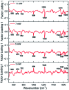
Hydroxyethyl starch (HES) is a commonly used intravenous fluid in hospital settings. The merits and demerits of its application is still a debatable topic. Investigating the interaction of external agents like intravenous fluids with blood cells is of great significance in clinical environments. Micro-Raman spectroscopy combined with an optical tweezers technique has been utilized for conducting systematic investigations of single live red blood cells (RBCs) under the influence of external stress agents. The present work deals with a detailed biophysical study on the response of human live red blood cells in hydroxyethyl starch using optical techniques. Morphological changes in red blood cells were monitored using quantitate phase imaging techniques. Micro-Raman studies suggest that there is a significant reduction in the oxy-haemoglobin level in red blood cells suspended in HES. The spectra recorded by using different probe laser powers has shown that the cells are more vulnerable in HES under the influence of externally induced stress than in blood plasma. In addition, the spectral results support the possibility of heme aggregation and membrane damage for red blood cells in HES under externally induced stress. Principle component analysis performed on the Raman spectra were able to effectively discriminate between red blood cells in HES and in blood plasma. The use of Raman tweezers can be highly beneficial in elucidating biochemical alterations happening in live, human red blood cell.
This journal is © The Royal Society of Chemistry.
Conflict of interest statement
The authors declare that they have no known competing financial interests or personal relationships that could have appeared to influence the work reported in this paper.
Figures


References
-
- Jenkins C. A.; Chandler S.; Jenkins R.; Thorne K.; Cunningham A.; Nelson K.; Still R.; Walters J.; Gwynn N.; Chea W., A New Method to Triage Colorectal Cancer Referrals in the UK Using Serum Raman Spectroscopy: A Prospective Cohort Study. 2020
-
- Correia N. A. Batista L. T. Nascimento R. J. Cangussú M. C. Crugeira P. J. Soares L. G. Silveira Jr L. Pinheiro A. L. Detection of prostate cancer by Raman spectroscopy: A multivariate study on patients with normal and altered PSA values. J. Photochem. Photobiol., B. 2020;204:111801. doi: 10.1016/j.jphotobiol.2020.111801. - DOI - PubMed
LinkOut - more resources
Full Text Sources
Other Literature Sources

