. 2020 Nov 4;10(66):40157-40170.
doi: 10.1039/d0ra06873j. eCollection 2020 Nov 2.

An antibacterial and injectable calcium phosphate scaffold delivering human periodontal ligament stem cells for bone tissue engineering

Affiliations

An antibacterial and injectable calcium phosphate scaffold delivering human periodontal ligament stem cells for bone tissue engineering

Hong Chen et al. RSC Adv. .

Abstract

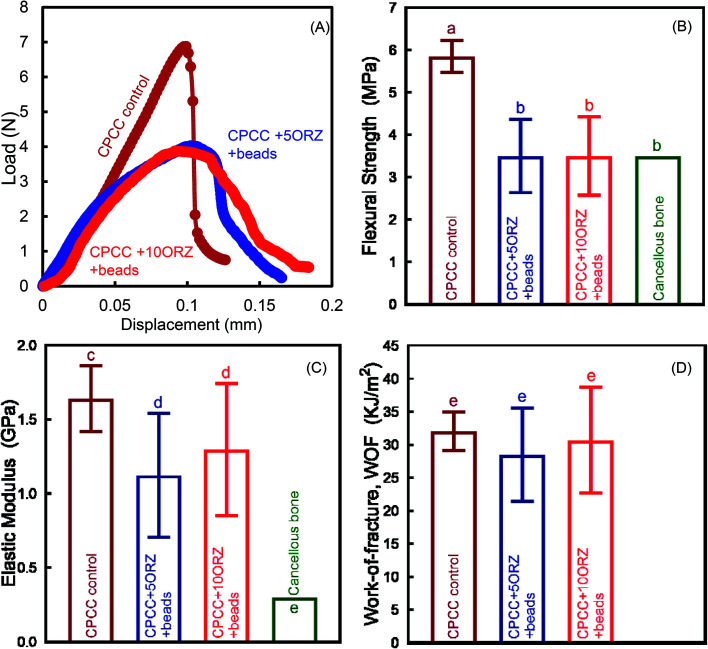
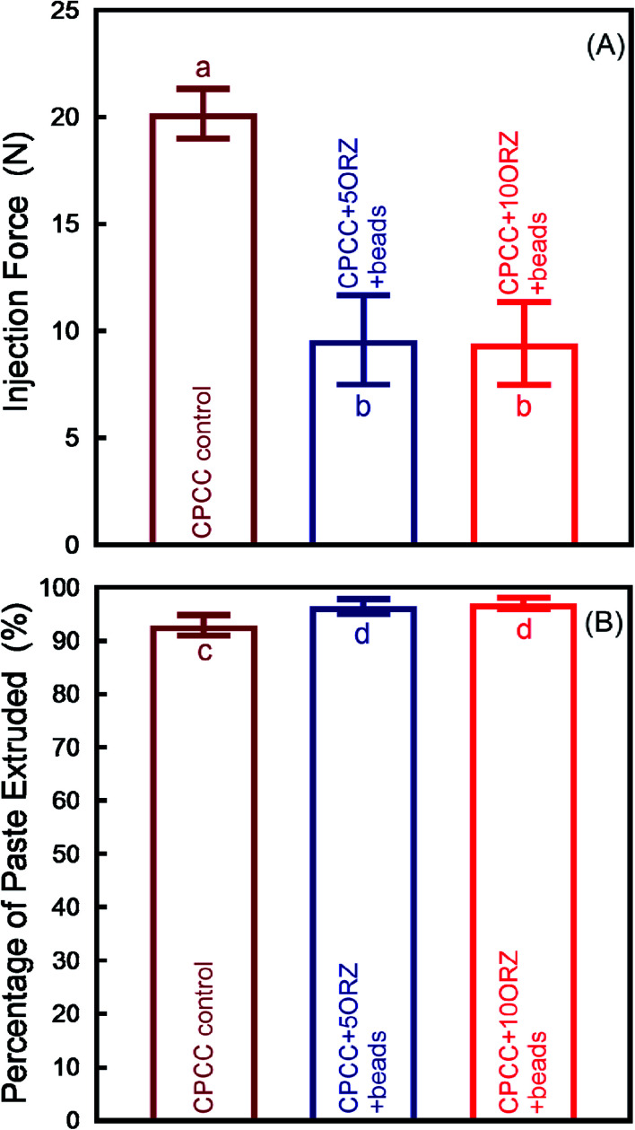
Osteomyelitis and post-operative infections are major problems in orthopedic, dental and craniofacial surgeries. It is highly desirable for a tissue engineering construct to kill bacteria, while simultaneously delivering stem cells and enhancing cell function and tissue regeneration. The objectives of this study were to: (1) develop a novel injectable calcium phosphate cement (CPC) scaffold containing antibiotic ornidazole (ORZ) while encapsulating human periodontal ligament stem cells (hPDLSCs), and (2) investigate the inhibition efficacy against Staphylococcus aureus (S. aureus) and the promotion of hPDLSC function for osteogenesis for the first time. ORZ was incorporated into a CPC-chitosan scaffold. hPDLSCs were encapsulated in alginate microbeads (denoted hPDLSCbeads). The ORZ-loaded CPCC+hPDLSCbeads scaffold was fully injectable, and had a flexural strength of 3.50 ± 0.92 MPa and an elastic modulus of 1.30 ± 0.45 GPa, matching those of natural cancellous bone. With 6 days of sustained ORZ release, the CPCC+10ORZ (10% ORZ) scaffold had strong antibacterial effects on S. aureus, with an inhibition zone of 12.47 ± 1.01 mm. No colonies were observed in the CPCC+10ORZ group from 3 to 7 days. ORZ-containing scaffolds were biocompatible with hPDLSCs. CPCC+10ORZ+hPDLSCbeads scaffold with osteogenic medium had 2.4-fold increase in alkaline phosphatase (ALP) activity and bone mineral synthesis by hPDLSCs, as compared to the control group (p < 0.05). In conclusion, the novel antibacterial construct with stem cell delivery had injectability, good strength, strong antibacterial effects and biocompatibility, supporting osteogenic differentiation and bone mineral synthesis of hPDLSCs. The injectable and mechanically-strong CPCC+10ORZ+hPDLSCbeads construct has great potential for treating bone infections and promoting bone regeneration.


Conflict of interest statement

There are no conflicts to declare.

Figures

Fig. 1
Fig. 1. Mechanical properties of CPC-chitosan scaffolds (mean ± sd; n = 6). Typical load–displacement curves (A); Flexural strength (B), elastic modulus (GPa) (C), and work-of-fracture (D). Values with dissimilar letters are significantly different from each other (p < 0.05).
Fig. 2
Fig. 2. Injectability of the CPC-chitosan scaffolds (mean ± sd; n = 6). Injection force (A), and percentage of paste extruded (B). Dissimilar letters indicate significantly different values (p < 0.05).
Fig. 3
Fig. 3. The drug release profile of ornidazole (ORZ) from CPC-chitosan scaffolds (mean ± sd; n = 6). After an initial burst of release of nearly 50% of the dose in 12 hour, the complete ORZ release was observed in the 3rd and 6th day for CPCC+5ORZ and CPCC+10ORZ, respectively.
Fig. 4
Fig. 4. Antimicrobial activity of CPC-chitosan scaffolds against S. aureus biofilm. The 24 hour images of the samples against S. aureus (A–D). The bacteriostatic rings of CPC-chitosan scaffolds against S. aureus (E) (mean ± sd; n = 6). Dissimilar letters indicate significantly different values (p < 0.05).
Fig. 5
Fig. 5. Representative live/dead staining images of 24 hour S. aureus biofilms (A–C). Time-kill curve showing log reduction against S. aureus (D) (mean ± sd; n = 6). Dissimilar letters indicate significantly different values (p < 0.05).
Fig. 6
Fig. 6. Cell viability assay of CPC-chitosan scaffolds with and without ornidazole (ORZ) addition (mean ± sd; n = 6). CPCC+5ORZ and CPCC+10ORZ groups had cell viability of above 90%. An increasing trend of the hPDLSC viability was shown in all groups over time.
Fig. 7
Fig. 7. Cell encapsulation and release from alginate microbeads (without the CPC-chitosan scaffolds) at 0 day (A, F and K). To examine microbeads degradation and cell release, 50% by volume of hPDLSC-encapsulating alginate microbeads was placed on the sterilized CPC disks. At 1 day, cell-encapsulating microbeads were collected and imaged (B, G and L). In the culture medium, the microbeads gradually degraded and the hPDLSCs (red arrows) started to be released from the microbeads at 4 days (C, H, M and P). More cells were released, and the contour of the microbeads became obscure as the alginate degraded from 4 to 14 days (D, E, I, J, N and O).
Fig. 8
Fig. 8. Live/dead images of hPDLSCs encapsulated in alginate microbeads in CPC-chitosan scaffolds (A–L). Live cells (green) were numerous and dead cells (red) were very few. Percentages of live cells (M) and CCK-8 from 1 to 14 days showed good cell viability and proliferation, and cell viability, which was not negatively affected by injection force and the delivery of ORZ (N) (mean ± sd; n = 6). Dissimilar letters indicate significantly different values (p < 0.05).
Fig. 9
Fig. 9. Osteogenic differentiation of hPDLSCs encapsulated in alginate microbeads in CPC-chitosan scaffolds. (A) Alkaline phosphatase (ALP) staining and quantitative analysis after 14 days of osteogenic induction (n = 6). (B) Alizarin Red staining (ARS) and quantitative calculation after 14 days of osteogenic induction (n = 6). Dissimilar letters indicate significantly different values (p < 0.05).

Similar articles

Cited by

References

    1. Moskowitz J. S. Blaisse M. R. Samuel R. E. Hsu H.-P. Harris M. B. Martin S. D. Lee J. C. Spector M. Hammond P. T. Biomaterials. 2010;31:6019–6030. doi: 10.1016/j.biomaterials.2010.04.011. - DOI - PMC - PubMed
    1. Jenkins A. Diep B. A. Mai T. T. Vo N. H. Warrener P. Suzich J. Stover C. K. Sellman B. R. mBio. 2015;6(1):e02272-14. doi: 10.1128/mBio.02272-14. - DOI - PMC - PubMed
    1. Malizos K. Sarma J. Seaton R. Militz M. Menichetti F. Riccio G. Gaudias J. Trostmann U. Pathan R. Hamed K. Eur. J. Clin. Microbiol. Infect. Dis. 2016;35:111–118. doi: 10.1007/s10096-015-2515-6. - DOI - PMC - PubMed
    1. Horst S. A. Hoerr V. Beineke A. Kreis C. Tuchscherr L. Kalinka J. Lehne S. Schleicher I. Köhler G. Fuchs T. Am. J. Pathol. 2012;181:1206–1214. doi: 10.1016/j.ajpath.2012.07.005. - DOI - PubMed
    1. Jayasree R. Kumar T. S. Perumal G. Doble M. Ceram. Int. 2018;44:9227–9235. doi: 10.1016/j.ceramint.2018.02.133. - DOI