Extracellular histones are clinically associated with primary graft dysfunction in human liver transplantation
- PMID: 35520915
- PMCID: PMC9062399
- DOI: 10.1039/c9ra00425d
Extracellular histones are clinically associated with primary graft dysfunction in human liver transplantation
Erratum in
-
Correction: Extracellular histones are clinically associated with primary graft dysfunction in human liver transplantation.RSC Adv. 2019 Aug 22;9(45):26435. doi: 10.1039/c9ra90059d. eCollection 2019 Aug 19. RSC Adv. 2019. PMID: 35531002 Free PMC article.
Abstract
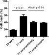
Extracellular histones have been involved in numerous inflammatory conditions such as ischemia/reperfusion (I/R) injury, trauma, and infection. There is growing evidence of I/R injury associated with primary graft dysfunction (PGD) following organ transplantation. Here we investigated whether extracellular histones are clinically involved with PGD in human liver transplantation. In total 58 patients undergoing liver transplantation were studied. We collected blood samples from the recipients before and serially after transplantation (24 h, 72 h). We measured extracellular histones, myeloperoxidase (MPO), S100A8/A9, and multiple inflammatory cytokines. Additionally, we exposed human L02 hepatocytes or U937 monocytic cells to the recipient's sera overnight, and assessed cellular viability and cytokine production respectively. Lastly, we assessed the effect of histone-targeted interventions by administration of heparin or an anti-histone antibody. It showed that extracellular histones increased immediately after transplantation, peaked within 24 hours and remained at high levels up to 72 hours (all p < 0.01). Notably, extracellular histone levels were significantly higher in recipients with PGD (n = 9) than recipients without PGD (n = 49, p = 0.004). Extracellular histones correlated positively with MPO, S100A8/A9 and most detected cytokines. Ex vivo analysis demonstrated that the patients' sera after graft markedly induced L02 cell death and caused profound cytokine production in cultured U937 cells, which could be abrogated by heparin or an anti-histone antibody. Collectively, extracellular histones were increased significantly after liver transplantation, which may contribute to the occurrence of PGD through direct cytotoxicity and enhancement of systemic inflammation. Targeting extracellular histones may provide a promising approach for preventing PGD or other complications in clinical practice.
This journal is © The Royal Society of Chemistry.
Conflict of interest statement
The authors declare that they have no conflict of interest.
Figures


Similar articles
-
Extracellular histones play a pathogenic role in primary graft dysfunction after human lung transplantation.RSC Adv. 2020 Mar 27;10(21):12485-12491. doi: 10.1039/d0ra00127a. eCollection 2020 Mar 24. RSC Adv. 2020. PMID: 35497627 Free PMC article.
-
Patients with HBV-related acute-on-chronic liver failure have increased concentrations of extracellular histones aggravating cellular damage and systemic inflammation.J Viral Hepat. 2017 Jan;24(1):59-67. doi: 10.1111/jvh.12612. Epub 2016 Sep 23. J Viral Hepat. 2017. PMID: 27660136
-
Circulating histones are major mediators of systemic inflammation and cellular injury in patients with acute liver failure.Cell Death Dis. 2016 Sep 29;7(9):e2391. doi: 10.1038/cddis.2016.303. Cell Death Dis. 2016. PMID: 27685635 Free PMC article.
-
Primary graft dysfunction after liver transplantation.Hepatobiliary Pancreat Dis Int. 2014 Apr;13(2):125-37. doi: 10.1016/s1499-3872(14)60023-0. Hepatobiliary Pancreat Dis Int. 2014. PMID: 24686540 Review.
-
Interventions for preventing thrombosis in solid organ transplant recipients.Cochrane Database Syst Rev. 2021 Mar 15;3(3):CD011557. doi: 10.1002/14651858.CD011557.pub2. Cochrane Database Syst Rev. 2021. PMID: 33720396 Free PMC article.
Cited by
-
Anti-DAMP therapies for acute inflammation.Front Immunol. 2025 May 8;16:1579954. doi: 10.3389/fimmu.2025.1579954. eCollection 2025. Front Immunol. 2025. PMID: 40406124 Free PMC article. Review.
-
Advancements in Predictive Tools for Primary Graft Dysfunction in Liver Transplantation: A Comprehensive Review.J Clin Med. 2024 Jun 27;13(13):3762. doi: 10.3390/jcm13133762. J Clin Med. 2024. PMID: 38999328 Free PMC article. Review.
-
Potential Use of Anti-Inflammatory Synthetic Heparan Sulfate to Attenuate Liver Damage.Biomedicines. 2020 Nov 16;8(11):503. doi: 10.3390/biomedicines8110503. Biomedicines. 2020. PMID: 33207634 Free PMC article. Review.
-
Liquid Liver Biopsy for Disease Diagnosis and Prognosis.J Clin Transl Hepatol. 2023 Dec 28;11(7):1520-1541. doi: 10.14218/JCTH.2023.00040. Epub 2023 Jul 27. J Clin Transl Hepatol. 2023. PMID: 38161500 Free PMC article. Review.
References
LinkOut - more resources
Full Text Sources
Research Materials
Miscellaneous

