

Frontiers in Lichen Planopilaris and Frontal Fibrosing Alopecia Research: Pathobiology Progress and Translational Horizons
- PMID: 35521043
- PMCID: PMC9062486
- DOI: 10.1016/j.xjidi.2022.100113
Frontiers in Lichen Planopilaris and Frontal Fibrosing Alopecia Research: Pathobiology Progress and Translational Horizons
Erratum in
-
Erratum.JID Innov. 2022 Aug 2;3(2):100149. doi: 10.1016/j.xjidi.2022.100149. eCollection 2023 Mar. JID Innov. 2022. PMID: 36909740 Free PMC article.
Abstract
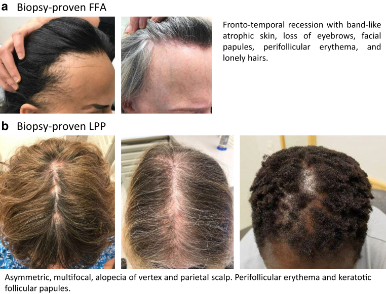
Lichen planopilaris (LPP) and frontal fibrosing alopecia (FFA) are primary, lymphocytic cicatricial hair loss disorders. These model epithelial stem cell (SC) diseases are thought to result from a CD8+ T-cell‒dominated immune attack on the hair follicle (HF) SC niche (bulge) after the latter has lost its immune privilege (IP) for as yet unknown reasons. This induces both apoptosis and pathological epithelial‒mesenchymal transition in epithelial SCs, thus depletes the bulge, causes fibrosis, and ultimately abrogates the HFs' capacity to regenerate. In this paper, we synthesize recent progress in LPP and FFA pathobiology research, integrate our limited current understanding of the roles that genetic, hormonal, environmental, and other factors may play, and define major open questions. We propose that LPP and FFA share a common initial pathobiology, which then bifurcates into two distinct clinical phenotypes, with macrophages possibly playing a key role in phenotype determination. As particularly promising translational research avenues toward direly needed progress in the management of these disfiguring, deeply distressful cicatricial alopecia variants, we advocate to focus on the development of bulge IP and epithelial SC protectants such as, for example, topically effective, HF‒penetrating and immunoinhibitory preparations that contain tacrolimus, peroxisome proliferator-activated receptor-γ, and/or CB1 agonists.
Keywords: 5ARI, 5α-reductase inhibitor; AA, alopecia areata; AGA, androgenetic alopecia; CRH, corticotropin-releasing hormone; EMT, epithelial‒mesenchymal transition; FFA, frontal fibrosing alopecia; HF, hair follicle; IP, immune privilege; K, keratin; KC, keratinocyte; LPP, lichen planopilaris; MAC, macrophage; MHC, major histocompatibility complex; PCA, primary cicatricial alopecia; PCP, personal care product; PPAR-γ, peroxisome proliferator–activated receptor-γ; SC, stem cell; SP, substance P; eHFSC, epithelial hair follicle stem cell; α-MSH, α-melanocyte-stimulating hormone.
© 2022 The Authors.
Figures


References
-
- Aldoori N., Dobson K., Holden C.R., McDonagh A.J., Harries M., Messenger A.G. Frontal fibrosing alopecia: possible association with leave-on facial skin care products and sunscreens; a questionnaire study. Br J Dermatol. 2016;175:762–767. - PubMed
-
- Arck P.C., Handjiski B., Hagen E., Joachim R., Klapp B.F., Paus R. Indications for a “brain-hair follicle axis (BHA)”: inhibition of keratinocyte proliferation and up-regulation of keratinocyte apoptosis in telogen hair follicles by stress and substance P. FASEB J. 2001;15:2536–2538. - PubMed
-
- Arck P.C., Handjiski B., Kuhlmei A., Peters E.M.J., Knackstedt M., Peter A., et al. Mast cell deficient and neurokinin-1 receptor knockout mice are protected from stress-induced hair growth inhibition. J Mol Med (Berl) 2005;83:386–396. - PubMed
-
- Atarguine H., Hocar O., Hamdaoui A., Akhdari N., Amal S. [Frontal fibrosing alopecia: report on three pediatric cases] Arch Pediatr. 2016;23:832–835. - PubMed
-
- Baibergenova A., Donovan J. Lichen planopilaris: update on pathogenesis and treatment. Skinmed. 2013;11:161–165. - PubMed
Publication types
LinkOut - more resources
Full Text Sources
Research Materials
Miscellaneous
