. 2022 Apr 16;25(5):104263.
doi: 10.1016/j.isci.2022.104263. eCollection 2022 May 20.

Organic acid cross-linked 3D printed cellulose nanocomposite bioscaffolds with controlled porosity, mechanical strength, and biocompatibility

Affiliations

Organic acid cross-linked 3D printed cellulose nanocomposite bioscaffolds with controlled porosity, mechanical strength, and biocompatibility

Andreja Dobaj Štiglic et al. iScience. .

Abstract

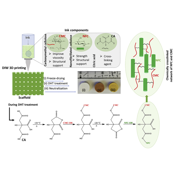
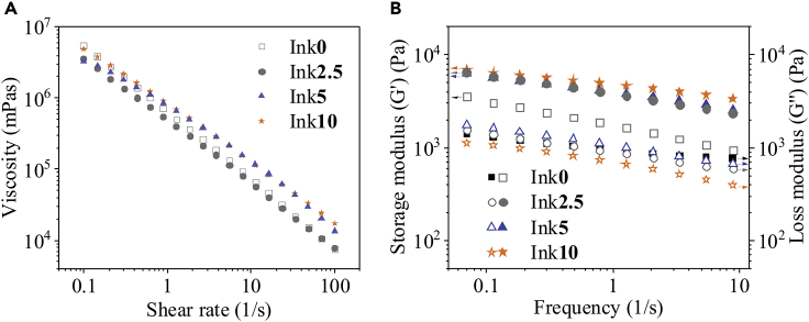
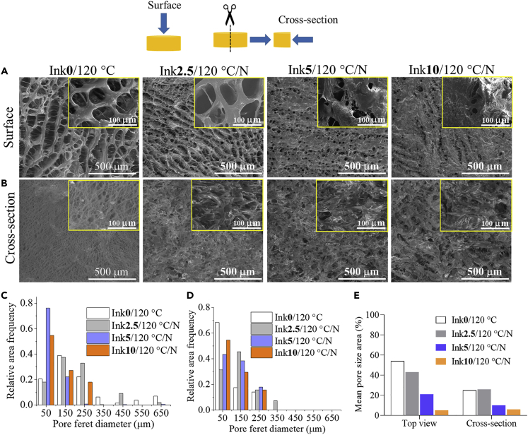
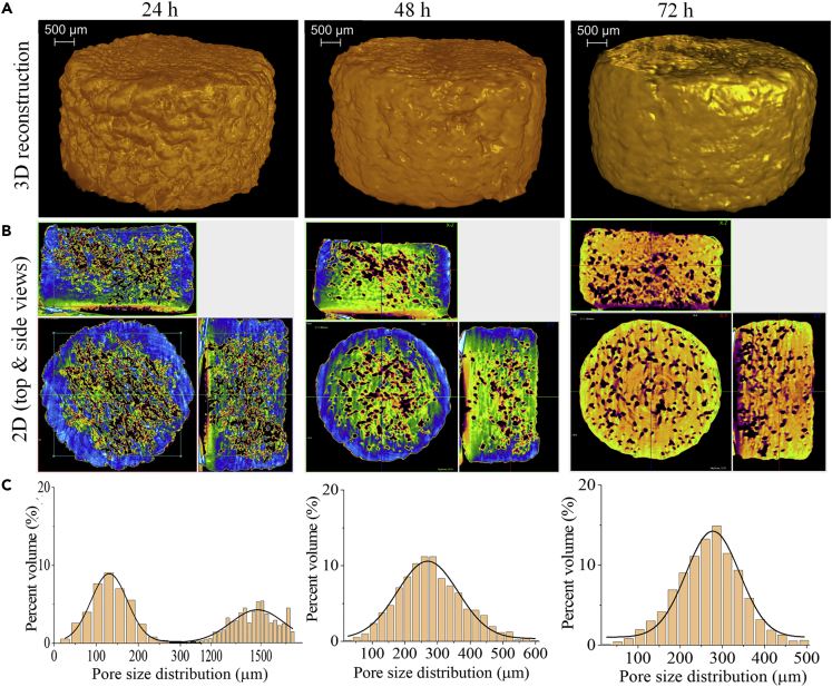
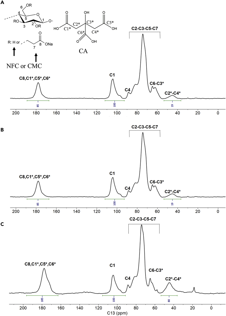
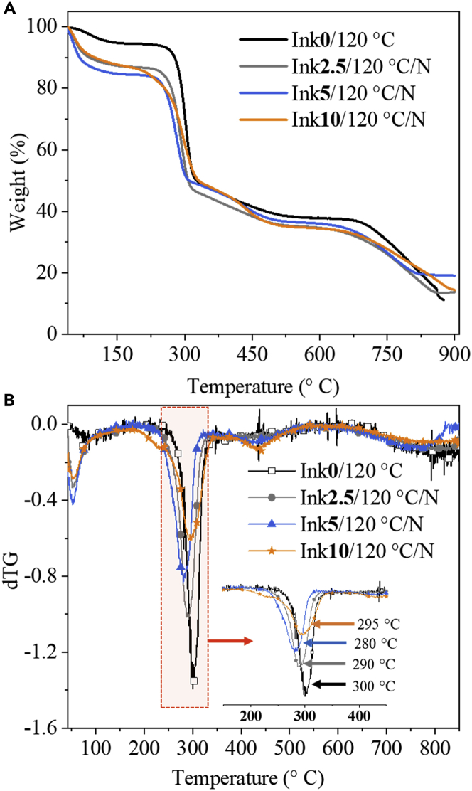
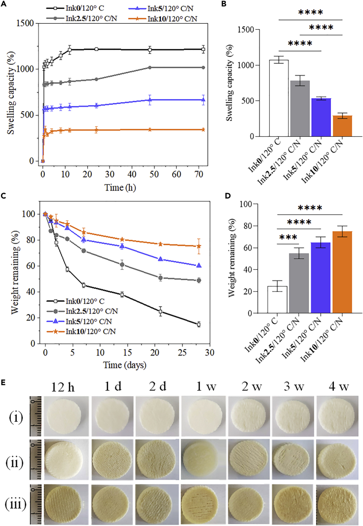
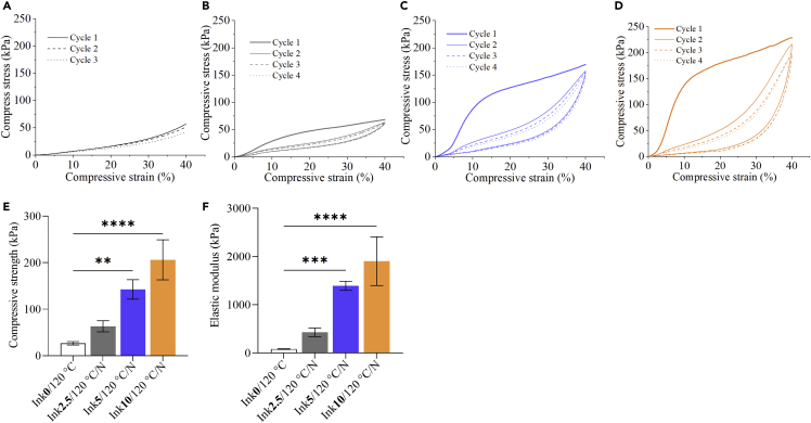
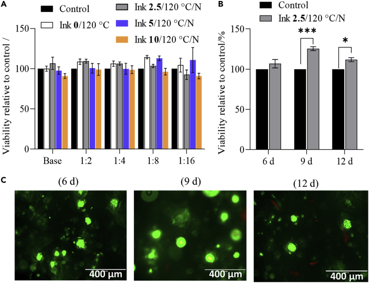
Herein, we fabricated chemically cross-linked polysaccharide-based three-dimensional (3D) porous scaffolds using an ink composed of nanofibrillated cellulose, carboxymethyl cellulose, and citric acid (CA), featuring strong shear thinning behavior and adequate printability. Scaffolds were produced by combining direct-ink-writing 3D printing, freeze-drying, and dehydrothermal heat-assisted cross-linking techniques. The last step induces a reaction of CA. Degree of cross-linking was controlled by varying the CA concentration (2.5-10.0 wt.%) to tune the structure, swelling, degradation, and surface properties (pores: 100-450 μm, porosity: 86%) of the scaffolds in the dry and hydrated states. Compressive strength, elastic modulus, and shape recovery of the cross-linked scaffolds increased significantly with increasing cross-linker concentration. Cross-linked scaffolds promoted clustered cell adhesion and showed no cytotoxic effects as determined by the viability assay and live/dead staining with human osteoblast cells. The proposed method can be extended to all polysaccharide-based materials to develop cell-friendly scaffolds suitable for tissue engineering applications.

Keywords: Biomaterials; Materials science; Tissue Engineering.


Conflict of interest statement

The authors declare no competing interests.

Figures

None
Graphical abstract
Figure 1
Figure 1
Rheological properties of the inks prepared with different amounts of citric acid concentrations (0 –10 wt.%) Viscosity curves (A) and frequency-dependency of the storage (symbols completely filled) and loss shear moduli (symbols not filled) (B) of the inks composed of nanofibrillated cellulose (NFC), carboxymethyl cellulose (CMC), and citric acid at various concentrations (0–10 wt.%).
Figure 2
Figure 2
Illustration of the design of citric acid cross-linked three-dimensional scaffolds of nanofibrillated cellulose and carboxymethyl cellulose via the combination of direct ink writing printing, freeze-drying, and dehydrothermal heat treatment
Figure 3
Figure 3
SEM morphology and pore size analysis (A) Top view (B) Cross-section (side view) of dry scaffolds cross-linked without (Ink0/120°C) and with different concentrations of citric acid (0 – 10 wt.%). Magnification in images is 100× and 1000x (insert). Mean Feret diameter (μm, C: surface and D: cross-section) and pore size area (%, E) of non-cross-linked and CA-cross-linked scaffolds.
Figure 4
Figure 4
Micro-CT analysis of the dry scaffolds (A) 3D reconstruction and (B) 2D top and side views and (C) pore size distribution of scaffolds before and after cross-linking with different concentrations of citric acid.
Figure 5
Figure 5
Micro-CT analysis of the wet scaffolds (A) 3D reconstruction and (B) 2D top and side views and (C) pore size distribution of scaffold Ink2.5/120°C/N stored in water at different time points.
Figure 6
Figure 6
13C-solid state NMR spectra of NFC/CMC scaffolds cross-linked with different amounts of citric acid (–2.5–10 wt.%) (A) Ink2.5/120°C/N. (B) Ink5/120°C/N. (C) Ink10/120°C/N.
Figure 7
Figure 7
Thermal stabilities of cross-linked and non-cross-linked scaffolds TGA (A) and dTG (B) curves of NFC/CMC scaffolds cross-linked with different citric acid concentrations.
Figure 8
Figure 8
Swelling and degradation of cross-linked and non-cross-linked scaffolds Swelling (A, B) and weight loss (C, D) of NFC/CMC scaffolds cross-linked with different citric acid concentrations. (E) Images of NFC/CMC scaffolds (i: Ink0/120°C, ii: Ink2.5/120°C/N and iii: Ink10/120°C/N) taken after the weight loss test at different times. Data analysis was done by one-ANOVA with the Dunnett test, and values are presented as ± SD; ∗∗∗p < 0.05, ∗∗∗∗p < 0.05 (compared to control Ink0/120°C).
Figure 9
Figure 9
Cyclic compression curves of the of NFC/CMC scaffolds cross-linked with different CA concentrations (A: Ink0/120°C, B: Ink2.5/120°C/N, C: Ink5/120°C/N, D: Ink10/120°C/N). Compressive strength (at 30% strain) (E) and elastic modulus (F) were extracted from compression tests in Figures 9A–9D. Data analysis was done by one-ANOVA with the Dunnett test, values are presented as ± SD; ∗∗p < 0.05, ∗∗∗∗p < 0.05 (compared to control Ink0/120°C).
Figure 10
Figure 10
Human osteoblast cell’s viability exposed to the prepared samples (A)MTT assay on sample extracts at different dilutions; experiments were performed for 24 h. (B)XTT results one the scaffold Ink 2.5/120°C/N tested at different time intervals. (C)fluorescence images of osteoblast cells cultured on scaffold Ink 2.5/120°C/N stained using the Live/dead assay after 6, 9ays and 12 days. Data analysis was done by one-student t-test (unpaired), values are presented as ± SD; ∗∗∗p < 0.05, ∗p < 0.05, (compared to control scaffold Ink 2.5/120°C/N, tested without cells).

Similar articles

Cited by

References

    1. Abbasi N., Hamlet S., Love R.M., Nguyen N.-T. Porous scaffolds for bone regeneration. J. Sci. Adv. Mater. Devices. 2020;5:1–9. doi: 10.1016/j.jsamd.2020.01.007. - DOI
    1. Bahrami N., Farzin A., Bayat F., Goodarzi A., Salehi M., Karimi R., Mohamadnia A., Parhiz A., Ai J. Optimization of 3D alginate scaffold properties with interconnected porosity using freeze-drying method for cartilage tissue engineering application. Arch. Neurosci. 2019;6 doi: 10.5812/ans.85122. - DOI
    1. Bartoš M., Suchý T., Foltán R. Note on the use of different approaches to determine the pore sizes of tissue engineering scaffolds: what do we measure? Biomed. Eng. Online. 2018;17:110. doi: 10.1186/s12938-018-0543-z. - DOI - PMC - PubMed
    1. Berillo D., Volkova N. Preparation and physicochemical characteristics of cryogel based on gelatin and oxidised dextran. J. Mater. Sci. 2014;49:4855–4868. doi: 10.1007/s10853-014-8186-3. - DOI
    1. Bose S., Vahabzadeh S., Bandyopadhyay A. Bone tissue engineering using 3D printing. Mater. Today. 2013;16:496–504. doi: 10.1016/j.mattod.2013.11.017. - DOI

LinkOut - more resources