. 2022;19(1):719-734.
doi: 10.1080/15476286.2022.2071517. Epub 2021 Dec 31.

Upon heat stress processing of ribosomal RNA precursors into mature rRNAs is compromised after cleavage at primary P site in Arabidopsis thalian a

Affiliations

Upon heat stress processing of ribosomal RNA precursors into mature rRNAs is compromised after cleavage at primary P site in Arabidopsis thalian a

T Darriere et al. RNA Biol. 2022.

Abstract

Transcription and processing of 45S rRNAs in the nucleolus are keystones of ribosome biogenesis. While these processes are severely impacted by stress conditions in multiple species, primarily upon heat exposure, we lack information about the molecular mechanisms allowing sessile organisms without a temperature-control system, like plants, to cope with such circumstances. We show that heat stress disturbs nucleolar structure, inhibits pre-rRNA processing and provokes imbalanced ribosome profiles in Arabidopsis thaliana plants. Notably, the accuracy of transcription initiation and cleavage at the primary P site in the 5'ETS (5' External Transcribed Spacer) are not affected but the levels of primary 45S and 35S transcripts are, respectively, increased and reduced. In contrast, precursors of 18S, 5.8S and 25S RNAs are rapidly undetectable upon heat stress. Remarkably, nucleolar structure, pre-rRNAs from major ITS1 processing pathway and ribosome profiles are restored after returning to optimal conditions, shedding light on the extreme plasticity of nucleolar functions in plant cells. Further genetic and molecular analysis to identify molecular clues implicated in these nucleolar responses indicate that cleavage rate at P site and nucleolin protein expression can act as a checkpoint control towards a productive pre-rRNA processing pathway.

Keywords: Arabidopsis; Nucleolus; heat stress; rRNA processing; ribosome.


Conflict of interest statement

No potential conflict of interest was reported by the author(s).

Figures

Figure 1.
Figure 1.
Plant growth and nucleolus organization in response to heat stress. A) Arabidopsis seedlings maintained at 22°C (control), heat treated (37°C for 5 h, 8 h, and 24 h) or recovered R22°C-24 h (24 h at 37°C and then transferred 24 h at 22°C). B) Nucleolus structures visualized by TEM at 22°C and 37°C. Class I, regular nucleoli without (A) or with (B and C) Nucleolar Cavities (NoC). Class II, partially disrupted nucleoli (D-F); Class III, open (G and H) and entirely disrupted (I) nucleoli. C) Bar graph distribution (%, from 116 total nucleoli) of class I (dark blue), II (light blue), and III (white) nucleoli in non-treated control (22°C), heat treated (37°C), and recovered (R22°C-24 h) seedlings.
Figure 2.
Figure 2.
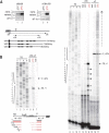
Expression of 3’ETS rDNA variants and analysis of TIS and P site in the 5’ETS of rRNA sequences under heat stress. A) RT-PCR using cDNAs prepared from non-treated (22°C, lanes 1 and 3) and heat treated (24 h, 37°C, lanes 2 and 4) plants. 3’ETS schematic representation below shows R1-4 repeat sequences, primers pairs o66/o36 and o108/o109, and expected amplification sequences. Amplification of eIF1α transcripts was performed to verify similar amounts of cDNA in each sample. B) Primer extension assays were performed on total RNAs from non-treated (22°C) and heat treated (37°C for 24 h) seedlings. Primers oTIS and oP detect respectively Transcription Initiation Site (TIS at +1) and the P cleavage site (at +1274). Reactions with oTIS+oP detects simultaneously TIS and P sites at 22°C and 37°C (lanes 5 and 6). Reactions with oTIS or oP only detects TIS and P sites at 22°C (lanes 13 and 16) or at 37°C (lanes 14 and 17). Lanes 1–4 and 8–11 are rDNA sequencing reactions with primer oTIS used to map transcription from TIS and rRNAs cleaved at P. Lanes 7, 12 and 15, mock control reactions using yeast tRNA. Schemes below show relative positions of primers oTIS and oP and the rRNA species detected at 22°C and 37°C.
Figure 3.
Figure 3.
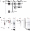
Pre-rRNA processing in response to heat stress. A) Scheme representing 45S rDNA and pre-RNA transcripts detected with probes hybridizing 5’ETS (p23), ITS1 (p43), ITS2 (p5), and 3’ETS (p6) sequences. rRNA precursors and fragments from major ITS1-first (black labelled) and minor 5’ETS-first (grey labelled) pathways are illustrated. B) Northern blot analysis of total RNAs from non-treated (even lanes) and heat treated at 37°C during 24 h (odd lanes) seedlings. A first membrane was hybridized with p23 and p5 (lanes 1–4) and a second one with p43 and p6 (lanes 7–10). Detected pre-rRNAs are labelled accordingly to previous reports using same probes. Pre-rRNAs detected upon heat stress are red labelled. Both membranes were stained with Gel Red to verify quality and amount of RNAs from samples at 22°C (lanes 5 and 11) and 37°C (lanes 6 and 12) and to localize the relative position of rRNAs 18S and 25S. The asterisk in lane 6 indicates a sporadic and unknown RNA species. RNA amounts for each sample were also verified by hybridization with p5S to detect 5S rRNA.
Figure 4.
Figure 4.
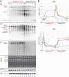
Kinetics of pre-rRNA processing throughout heat stress and recovery conditions. A) Northern blot analyses of total RNAs from non-treated (lane 1), heat treated (lanes 2–8), and recovered (lanes 9–13) seedlings. The same membrane was hybridized with p23, p43, and p6 probes, and similar RNA amounts for each sample were verified by Gel Red staining and hybridization with p5S and p25S to detect respectively 5S and 25S rRNAs. Pre-rRNAs detected upon heat stress are red labelled. B) Graphs show accumulation of pre-rRNAs 45S and 35S during heat (red underlined) and recovery (black underlined) periods. Relative amounts of each pre-rRNA detected with p23, p43 and p6 in each lane were determined, normalized to 5S rRNA signals (Table S5) and represented in blue, Orange and grey respectively.
Figure 5.
Figure 5.
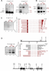
cRT-PCR amplifications to detect rRNA transcripts and products upon heat stress. The upper schemes represent a 45S rRNA sequences to show positions of primers used for cRT-PCR amplifications. cRT-PCR was performed on circularized RNAs from non-treated (22°C) or heat treated (37°C for 5 h or 24 h) seedlings with primers rt1, rtt3, or p31 for RT, and A) primers pairs r5+ r8 and r5+ r7, B) r10+ r2, C) r5+ r9, and D) p32+ p33 for PCR amplifications. Circular RT-PCR amplification products detected at 22°C and 37°C and identified by sequencing are respectively represented in black and in red. cRT-PCR products deduced by sizes are labelled in grey. The products sequenced after cRT-PCR reactions with rt1/r5+ r9 and p31/p32+ p33 are represented. Given that the primers used are divergent, part of the cDNA obtained from each circularized rRNA transcript (dotted lines) is absent from the final circular RT-PCR products. For each rRNA transcript, the 5’ and 3’ end positions are indicated. The number of sequenced clones are indicated on the left between brackets.
Figure 6.
Figure 6.
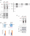
Pre-RNA processing in xrn2-3 and nuc1-2 in response to heat-stress. A) Northern blots analysis of total RNAs from Col-0 Arabidopsis WT and xrn2-3 seedlings non-treated (22°C, lanes 1 and 3) and heat-treated (37°C for 24 h, lanes 2 and 4) using probe p23. pre-rRNA transcripts 35S, P-A3 and fragments P-P’/P1 detected in WT are indicated in black and 35S*, 5’ETS-A3 and 5’ETS-P1 specifically detected in xrn2-3 [11] are in purple. The rRNA transcripts detected upon heat stress are red labelled: 45S, arrows, and arrowheads. Similar amounts of RNAs in each sample were verified by Gel Red staining. B) Western blot analysis of total protein extracts from two biological replicates of non-treated (lanes 1) and heat treated (37°C) for 24 h (lanes 2) seedlings. α-NUC1 antibody detects NUC1 protein (arrow) and two unspecific protein bands (asterisks, and see Figure S6B). Actin and Ponceau S are used as loading controls. The bar graphs show the relative amount of NUC1 in each Western blot. α-NUC1 and α-actin signals were quantified with ImageJ, and the amount of NUC1 normalized to actin values (Table S3). C) Northern blots analysis of total RNAs from Arabidopsis WT and nuc1-2 seedlings in non-treated (22°C, lane 1 and 3) and heat-treated (37°C for 24 h, lanes 2 and 4) using probe p23. Pre-rRNAs detected specifically in the heat stressed plant samples are indicated in red: 45S, arrows, and arrowheads. Similar amounts of RNAs in each sample were verified by Gel Red staining and hybridization with p5S to detect 5S rRNA.
Figure 7.
Figure 7.
Ribosome profiles and LC-MS/MS analysis on ribosome subunits in response to heat stress. A) Extracts from two experimental replicates (black and grey) of non-treated (22°C), heat treated (37°C for 5 h and 24 h), and recovered (37°C for 24 h then 22°C for 5 h and 24 h) seedlings were fractionated through 15–60% sucrose gradients. The percentage of the full scale of absorbance was monitored at 254 nm. Peaks corresponding to 40S and 60S-80S ribosomal subunits/monosomes are indicated and controlled by the presence of mature 18S and/or 25S rRNAs (Figure S7). The ratios of the 60–80S over the 40S were calculated for each replicate at each temperature, and the average Ratio (R60-80/40S) for each condition is indicated below. B) LC-MS/MS of ribosome subunits 40S and 60S-80S from sucrose gradient from non-treated (22°C) and 6 h, 37°C heat treated plants (Figure S7). Left Panel, relative amount (in %) of RPS, RPL and RPP in the 40S+60S+80S peaks at 22°C (fractions 8–11) and 37°C (fractions 9–12). Right panel, relative amounts of RPS, RPL and RPP in the 40S at 22°C (fractions 8 + 9) and (fractions 9–10) at 37°C or in the 60S-80S at 22°C (fractions 10 + 11) and at 37°C (fractions 11–12).

Similar articles

Cited by

References

    1. Henras AK, Plisson-Chastang C, O’Donohue MF, et al. An overview of pre-ribosomal RNA processing in eukaryotes. Wiley Interdiscip Rev RNA. 2015;6(2):225–242. - PMC - PubMed
    1. Sharma S, Lafontaine DL.. ‘view from a bridge’: a new perspective on eukaryotic rRNA base modification. Trends Biochem Sci. 2015;40(10):560–575. - PubMed
    1. Sloan KE, Warda AS, Sharma S, et al. Tuning the ribosome: the influence of rRNA modification on eukaryotic ribosome biogenesis and function. RNA Biol. 2017;14(9):1138–1152. - PMC - PubMed
    1. Tomecki R, Sikorski PJ, Zakrzewska-Placzek M. Comparison of preribosomal RNA processing pathways in yeast, plant and human cells - focus on coordinated action of endo- and exoribonucleases. FEBS Lett. 2017;591(13):1801–1850. - PubMed
    1. Saez-Vasquez J, Caparros-Ruiz D, Barneche F, et al. A plant snoRNP complex containing snoRNAs, fibrillarin, and nucleolin-like proteins is competent for both rRNA gene binding and pre-rRNA processing in vitro. Mol Cell Biol. 2004;24(16):7284–7297. - PMC - PubMed

Publication types