. 2022 Jan 1:47:bjac009.
doi: 10.1093/chemse/bjac009.

Target-specific projections of amygdala somatostatin-expressing neurons to the hypothalamus and brainstem

Affiliations

Target-specific projections of amygdala somatostatin-expressing neurons to the hypothalamus and brainstem

Jane J Bartonjo et al. Chem Senses. .

Abstract

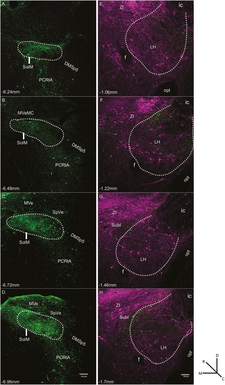
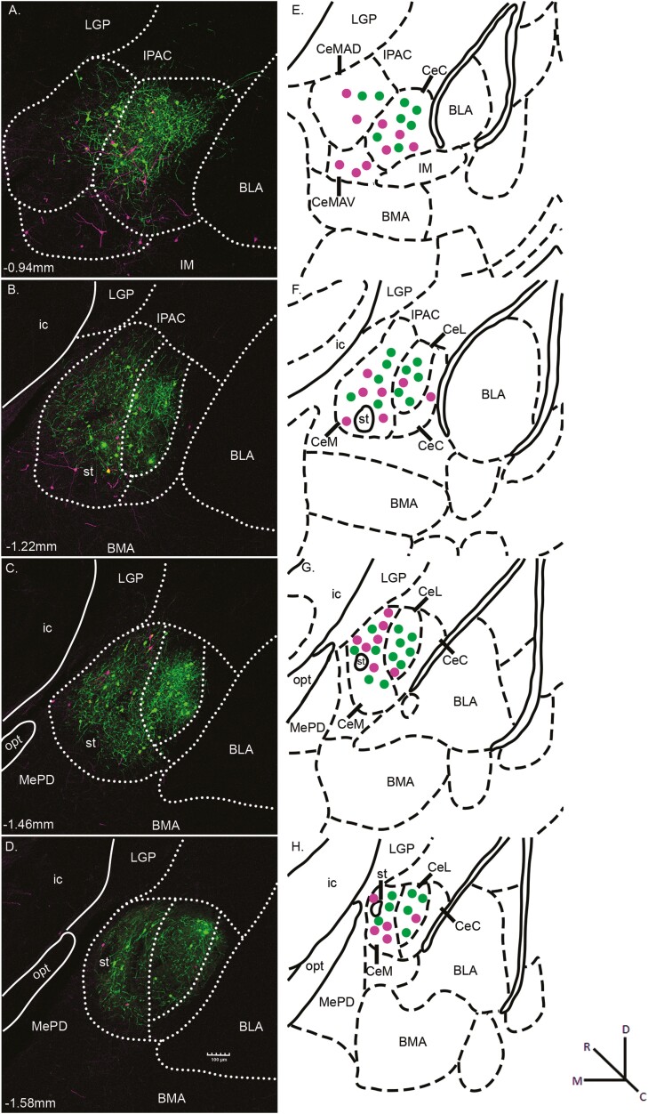
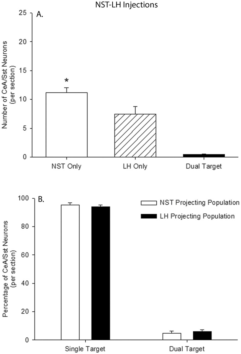
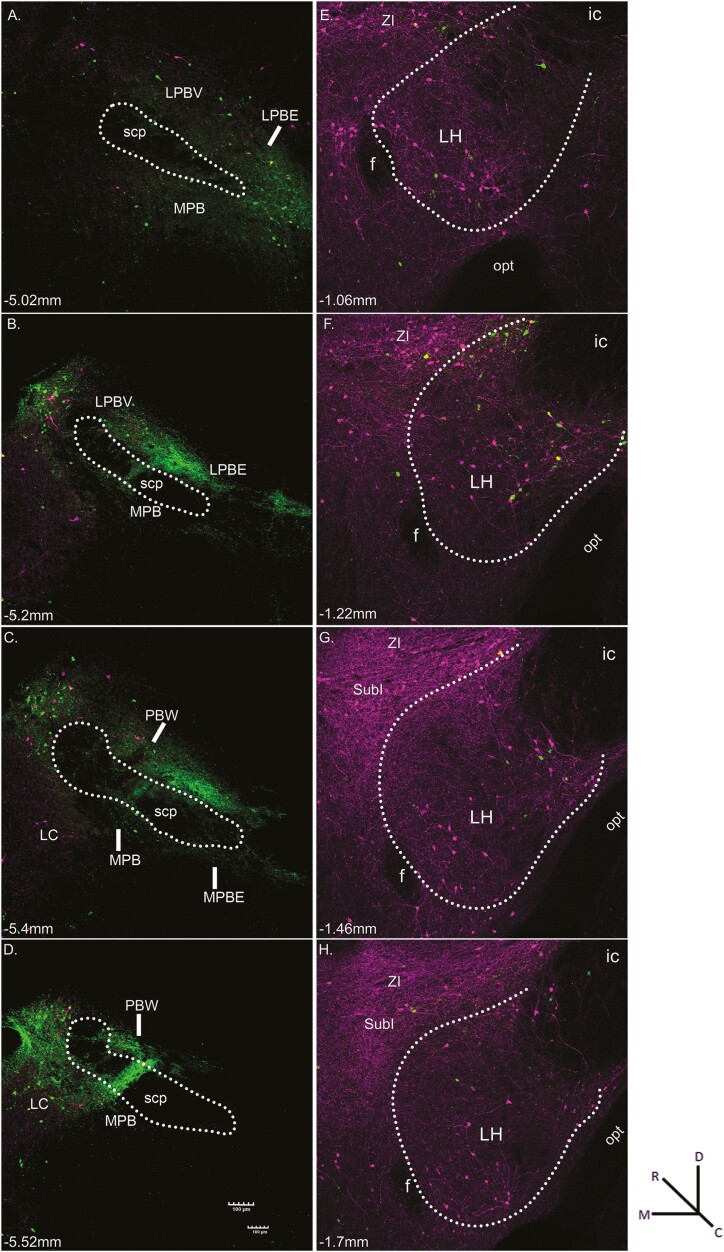
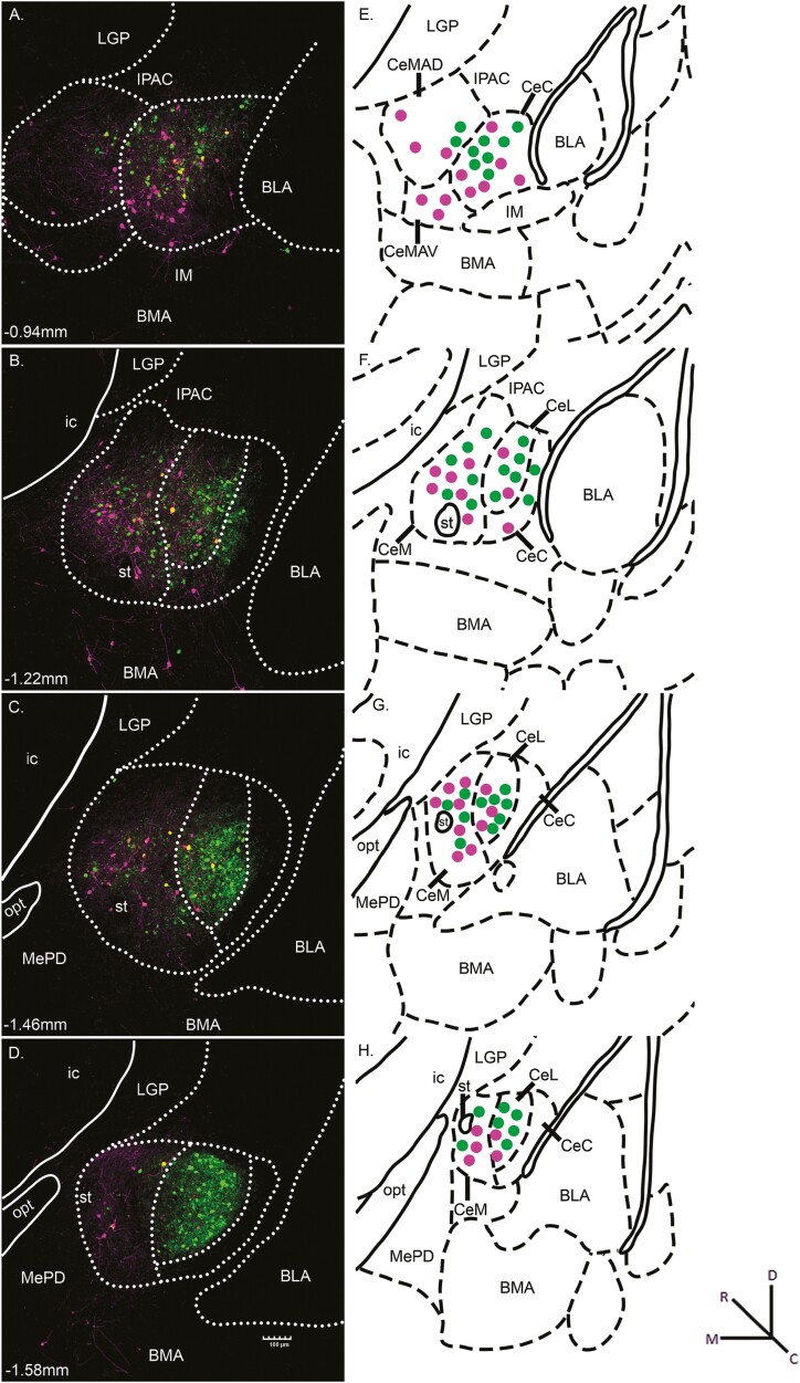
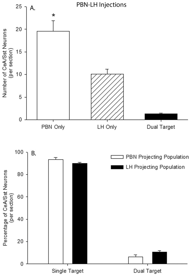
Somatostatin neurons in the central nucleus of the amygdala (CeA/Sst) can be parsed into subpopulations that project either to the nucleus of the solitary tract (NST) or parabrachial nucleus (PBN). We have shown recently that inhibition of CeA/Sst-to-NST neurons increased the ingestion of a normally aversive taste stimulus, quinine HCl (QHCl). Because the CeA innervates other forebrain areas such as the lateral hypothalamus (LH) that also sends axonal projections to the NST, the effects on QHCl intake could be, in part, the result of CeA modulation of LH-to-NST neurons. To address these issues, the present study investigated whether CeA/Sst-to-NST neurons are distinct from CeA/Sst-to-LH neurons. For comparison purposes, additional experiments assessed divergent innervation of the LH by CeA/Sst-to-PBN neurons. In Sst-cre mice, two different retrograde transported flox viruses were injected into the NST and the ipsilateral LH or PBN and ipsilateral LH. The results showed that 90% or more of retrograde-labeled CeA/Sst neurons project either to the LH, NST, or PBN. Separate populations of CeA/Sst neurons projecting to these different regions suggest a highly heterogeneous population in terms of synaptic target and likely function.

Keywords: NST; PBN; amygdala; herpes simplex virus; hypothalamus; taste.


Figures

Fig. 1.
Fig. 1.
Representative fluorescent images resulting from the injection of HSV-Ef1α-DIO-EYFP into the NST (A-D, green) and HSV-Ef1α-DIO-mCherry into the LH (E-H, magenta) of an Sst-cre mouse. Sections are arranged from rostral (top) to caudal (bottom). White dots outline the approximate boundaries of the NST and the LH. Within the NST, green fluorescence indicates Sst-expressing neurons that project locally within the NST. Within the LH, the magenta fluorescent cells indicate Sst-expressing interneurons, while those in surrounding areas such as ZI and SubI indicate Sst neurons that project to the LH. The green fluorescent cells represent Sst-positive LH-to-NST projection neurons, while the green fluorescent fibers likely represent retrograde-labeled axons from higher structures. The approximate level relative to bregma is shown at the bottom left of each photomicrograph (Paxinos and Franklin 2001). Magnification was 10× (0.85 pixels/μm). Abbreviations: DMsp5, dorsomedial spinal trigeminal nucleus; anterior part; f, fornix; ic, internal capsule; LH, lateral hypothalamus; MVe, medial vestibular nucleus; MVeMC, medial vestibular nucleus, magnocellular part; opt, optic tract; PCRtA, parvicellular reticular nucleus, alpha part; SolM, nucleus of the solitary tract, medial part; SpVe, spinal vestibular nucleus; SubI, substantia innominata; ZI, zona incerta.
Fig. 2.
Fig. 2.
(A-D) Representative photomicrographs of CeA/Sst neurons projecting to the NST (green) and LH (magenta) from HSV injections depicted in Fig. 1. Yellow fluorescence indicates Sst neurons that project both to the NST and LH. The white dotted lines outline the approximate boundaries of the CeC, CeL, and CeM divisions of the CeA. The approximate levels relative to bregma are indicated at the bottom left corner of each photomicrograph. Magnification of fluorescent images was 10× (0.85 pixels/μm). (E-H) Corresponding diagrams labeled with amygdala subnuclei as defined in Paxinos and Franklin (2001). The general location of retrograde labeled neurons is represented by green (NST projecting) and magenta (LH projecting) dots. Abbreviations: BLA, basolateral amygdaloid nucleus, anterior part; BMA, basomedial amygdaloid nucleus, anterior part; CeC, central amygdaloid nucleus, capsular part; CeL, central amygdaloid nucleus, lateral division; CeM, central amygdaloid nucleus, medial division; CeMAD, central amygdaloid nucleus, medial division, anterodorsal part; CeMAV, central amygdaloid nucleus, medial division, anteroventral part; IM, intercalated amygdaloid nucleus, main part; ic, internal capsule; IPAC, interstitial nucleus of the posterior limb of the anterior commissure; LGP, lateral globus pallidus; MePD, medial amygdaloid nucleus, posterodorsal part; opt, optic tract; st, stria terminalis.
Fig. 3.
Fig. 3.
(A) The per section average of retrograde labeled Sst neurons in the CeA following HSV injections into the NST and LH of Sst-cre mice. Open bar represents the average number of Sst cells that are only projected to the NST, cross-hatched bar Sst cells that are only projected to the LH, and filled bar Sst cells that are projected both to the NST and LH. *, NST Only >LH Only. (B) The mean percentage of single- and double-labeled Sst neurons in the CeA following HSV injections into the NST (open bars) and LH (filled bars).
Fig. 4.
Fig. 4.
Representative fluorescent images resulting from the injection of HSV-Ef1α-DIO-EYFP into the PBN (A-D, green) and HSV-Ef1α-DIO-mCherry into the LH (E-H, magenta) of an Sst-cre mouse. Sections are arranged from rostral (top) to caudal (bottom). White dots outline the approximate boundaries of the LH and superior cerebellar peduncle (scp) in the PBN. Within the PBN, the green fluorescent cells indicate Sst-expressing interneurons, while those in surrounding areas such as LC indicate Sst neurons that project to the PBN. The magenta fluorescent cells represent Sst-positive PBN-to-LH projection neurons. Within the LH, the magenta fluorescent cells indicate Sst-expressing interneurons, while those in surrounding areas such as ZI and SubI indicate Sst neurons that project to the LH. The green fluorescent cells represent Sst-positive LH-to-PBN projection neurons, while the green fluorescent fibers likely represent retrograde-labeled axons from higher structures. The approximate level relative to bregma is shown at the bottom left of each photomicrograph. Magnification was 10 × (0.85 pixels/micron). Abbreviations: f, fornix; ic, internal capsule; LC, locus coeruleus; LH, lateral hypothalamus; LPBE, lateral parabrachial nucleus, external part; LPBV, lateral parabrachial nucleus, ventral part; MPB, medial parabrachial nucleus; MPBe, medial parabrachial nucleus, external part; opt, optic tract; PBW, parabrachial nucleus, waist part; scp; superior cerebellar peduncle; SubI, substantia innominata; ZI, zona incerta.
Fig. 5.
Fig. 5.
(A-D) Representative photomicrographs of CeA/Sst neurons projecting to the PBN (green) and LH (magenta) from HSV injections depicted in Fig. 4. Yellow fluorescence indicates Sst neurons that project both to the PBN and LH. The white dotted lines outline the approximate boundaries of the CeC, CeL, and CeM divisions of the CeA. The approximate levels relative to bregma are indicated at the bottom left corner of each photomicrograph. Magnification of fluorescent images was 10 × (0.85 pixels/μm). (E-H) Corresponding diagrams labeled with amygdala subnuclei as defined in Paxinos and Franklin (2001). The general location of retrograde labeled neurons is represented by green (PBN projecting) and magenta (LH projecting) dots. Abbreviations: BLA, basolateral amygdaloid nucleus, anterior part; BMA, basomedial amygdaloid nucleus, anterior part; CeC, central amygdaloid nucleus, capsular part; CeL, central amygdaloid nucleus, lateral division; CeM, central amygdaloid nucleus, medial division; CeMAD, central amygdaloid nucleus, medial division, anterodorsal part; CeMAV, central amygdaloid nucleus, medial division, anteroventral part; IM, intercalated amygdaloid nucleus, main part; ic, internal capsule; IPAC, interstitial nucleus of the posterior limb of the anterior commissure; LGP, lateral globus pallidus; MePD, medial amygdaloid nucleus, posterodorsal part; opt, optic tract; st, stria terminalis.
Fig. 6.
Fig. 6.
(A) The per section average of retrograde labeled Sst neurons in the CeA following HSV injections into the PBN and LH of Sst-cre mice. Open bar represents the average number of Sst cells that are only projected to the PBN, cross-hatched bar Sst cells that are only projected to the LH, and filled bar Sst cells that are projected both to the PBN and LH. *, PBN Only > LH Only. (B) The mean percentage of single- and double-labeled Sst neurons in the CeA following HSV injections into the PBN (open bars) and LH (filled bars).

Similar articles

Cited by

References

    1. Balaban CD, Beryozkin G.. Vestibular nucleus projections to nucleus tractus solitarius and the dorsal motor nucleus of the vagus nerve: potential substrates for vestibulo-autonomic interactions. Exp Brain Res. 1994;98:200–212. - PubMed
    1. Barbier M, Fellmann D, Risold PY.. Morphofunctional organization of the connections from the medial and intermediate parts of the central nucleus of the amygdala into distinct divisions of the lateral hypothalamic area in the rat. Front Neurol. 2018;9:688. - PMC - PubMed
    1. Bartonjo J, Masterson S, St. John SJ, Lundy R.. Perturbation of amygdala/somatostatin-nucleus of the solitary tract projections reduces sensitivity to quinine in a brief-access test. Brain Res. 2022;1783:147838. - PMC - PubMed
    1. Bartonjo JJ, Lundy RF.. Distinct populations of amygdala somatostatin-expressing neurons project to the nucleus of the solitary tract and parabrachial nucleus. Chem Senses. 2020;45:687–698. - PMC - PubMed
    1. Beckman ME, Whitehead MC.. Intramedullary connections of the rostral nucleus of the solitary tract in the hamster. Brain Res. 1991;557:265–279. - PubMed

Publication types