Clinical Trial
. 2022 Jul 6;12(7):1656-1675.
doi: 10.1158/2159-8290.CD-21-1434.

Temozolomide Treatment Alters Mismatch Repair and Boosts Mutational Burden in Tumor and Blood of Colorectal Cancer Patients

Affiliations
Clinical Trial

Temozolomide Treatment Alters Mismatch Repair and Boosts Mutational Burden in Tumor and Blood of Colorectal Cancer Patients

Giovanni Crisafulli et al. Cancer Discov. .

Abstract

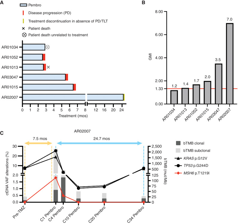
The majority of metastatic colorectal cancers (mCRC) are mismatch repair (MMR) proficient and unresponsive to immunotherapy, whereas MMR-deficient (MMRd) tumors often respond to immune-checkpoint blockade. We previously reported that the treatment of colorectal cancer preclinical models with temozolomide (TMZ) leads to MMR deficiency, increased tumor mutational burden (TMB), and sensitization to immunotherapy. To clinically translate these findings, we designed the ARETHUSA clinical trial whereby O6-methylguanine-DNA-methyltransferase (MGMT)-deficient, MMR-proficient, RAS-mutant mCRC patients received priming therapy with TMZ. Analysis of tissue biopsies and circulating tumor DNA (ctDNA) revealed the emergence of a distinct mutational signature and increased TMB after TMZ treatment. Multiple alterations in the nucleotide context favored by the TMZ signature emerged in MMR genes, and the p.T1219I MSH6 variant was detected in ctDNA and tissue of 94% (16/17) of the cases. A subset of patients whose tumors displayed the MSH6 mutation, the TMZ mutational signature, and increased TMB achieved disease stabilization upon pembrolizumab treatment.

Significance: MMR-proficient mCRCs are unresponsive to immunotherapy. We provide the proof of concept that inactivation of MMR genes can be achieved pharmacologically with TMZ and molecularly monitored in the tissue and blood of patients with mCRC. This strategy deserves additional evaluation in mCRC patients whose tumors are no longer responsive to standard-of-care treatments. See related commentary by Willis and Overman, p. 1612. This article is highlighted in the In This Issue feature, p. 1599.


Figures

Figure 1. The ARETHUSA trial. Graphical description of the ARETHUSA trial. (MGMT)-deficient RAS-mutant, and MMR-proficient mCRC patients received priming therapy with TMZ. A post-TMZ threshold of 20 mutations/Mb was required to access the immunotherapy phase delivering pembrolizumab every 3 weeks. MMRp, mismatch-repair proficient; MSS, microsatellite stable; IHC, immunohistochemistry; MB, methyl-BEAMing; TMZ, temozolomide; LB, liquid biopsy; WES, whole-exome sequencing; TMB, tumor mutational burden; PD, progressive disease; Pembro, pembrolizumab; mut, mutations; Mb, megabases.
Figure 1.
The ARETHUSA trial. Graphical description of the ARETHUSA trial. MGMT-deficient, RAS-mutant, and MMRp mCRC patients received priming therapy with TMZ. A post-TMZ TMB threshold of 20 mutations/Mb was required to access the immunotherapy phase delivering pembrolizumab every 3 weeks. IHC, immunohistochemistry; LB, liquid biopsy; MB, methyl-BEAMing; q28, every 28 days; q3w, every 3 weeks; Pembro, pembrolizumab; WES, whole exome sequencing.
Figure 2. Clinical response to TMZ in ARETHUSA patients. Swimmer plot of clinical time on treatment in the TMZ priming phase: 27 patients were treated with TMZ monotherapy until clinical or radiologic (based on RECIST 1.1 criteria) disease progression. Three patients AR02005, AR02007, and AR02011 were treated with TMZ-based regimens and enrolled in ARETHUSA according to protocol violation. Post-TMZ tissues for TMB evaluation were collected in 21 patients, 9 cases were excluded due to the clinical condition worsening. TMZ, temozolomide; TEMIRI, TMZ + irinotecan combination; CAPTEM, capecitabine + TMZ combination; mos, months of treatment.
Figure 2.
Clinical response to TMZ in ARETHUSA patients. Swimmer plot of clinical time on treatment in the TMZ priming phase: 27 patients were treated with TMZ monotherapy until clinical or radiologic (based on RECIST 1.1 criteria) disease progression. Three patients AR02005, AR02007, and AR02011 were treated with TMZ-based regimens and enrolled in ARETHUSA according to protocol violation. Post-TMZ tissues for TMB evaluation were collected in 21 patients; 9 cases were excluded due to the clinical condition worsening. CAPTEM, capecitabine + TMZ combination; mos, months of treatment; TEMIRI, TMZ + irinotecan combination.
Figure 3. Mutational signature and TMB analysis in biopsies after TMZ treatment. A and B, Mutational signature analysis measuring the impact of TMZ priming on tissue biopsies assessed by NGS. Patients were classified in three subtypes A, B1, and B2 based on the score of the mutational signature 11 and TMB value. In A, only clonal mutations (adjusted fractional abundance ≥ 10%) were used to generate the heatmap. In five cases (AR02071, AR01032, AR01014, AR01069, and AR02064), the number of mutations was not sufficient to properly perform mutational analysis (cosine similarity lower than 0.9) and these five samples were excluded. In B, all mutations (adjusted fractional abundance ≥ 1%) were considered for heatmap generation. C, TMB expressed as mut/MB after the priming phase for the three groups of patients. The relative contribution of clonal (yellow) and subclonal (blue) alterations to TMB is listed for each patient. Inset, Positive linear correlation between mutations induced by signature 11 (TMZ) normalized for megabases and temozolomide cycles of treatment. Spearman rank correlation is listed (P = 2.535e−5 and cor = 0.7847). D, TMB expressed as mutations/megabases after the priming phase for the three groups of patients. The relative contribution of SNVs and indels to TMB is listed for each patient. E, The best response to TMZ treatment is also reported for each patient. Subtype A (yellow): patients with no molecular evidence of TMZ treatment. Subtype B1 (blue): patients with subclonal molecular evidence of TMZ treatment. Subtype B2 (green): patients with clonal molecular evidence of TMZ treatment. TMZ, temozolomide; TMB, tumor mutational burden; Mut/MB, mutations for megabases; Sig, signature, SNV, single-nucleotide variant; INDEL, insertions and deletions; SD, stable disease; PD, progressive disease.
Figure 3.
Mutational signature and TMB analysis in biopsies after TMZ treatment. A and B, Mutational signature analysis measuring the impact of TMZ priming on tissue biopsies assessed by next-generation sequencing. Patients were classified in three subtypes—A, B1, and B2—based on the score of the mutational signature 11 and TMB value. In A, only clonal mutations (adjusted fractional abundance ≥10%) were used to generate the heat map. In five cases (AR02071, AR01032, AR01014, AR01069, and AR02064), the number of mutations was not sufficient to properly perform mutational analysis (cosine similarity lower than 0.9) and these five samples were excluded. In B, all mutations (adjusted fractional abundance ≥1%) were considered for heat map generation. C, TMB expressed as mut/Mb after the priming phase for the three groups of patients. The relative contribution of clonal (yellow) and subclonal (blue) alterations to TMB is listed for each patient. Inset, positive linear correlation between mutations induced by signature 11 (TMZ) normalized for megabases and TMZ cycles of treatment. Spearman rank correlation is listed (P = 2.535e−5 and R = 0.7847). D, TMB expressed as mut/Mb after the priming phase for the three groups of patients. The relative contribution of SNVs and indels to TMB is listed for each patient. E, The best response to TMZ treatment is also reported for each patient. Subtype A (yellow): patients with no molecular evidence of TMZ treatment. Subtype B1 (blue): patients with subclonal molecular evidence of TMZ treatment. Subtype B2 (green): patients with clonal molecular evidence of TMZ treatment. Sig, signature.
Figure 4. Comparison of signature and TMB analysis of tissue/blood samples before and after TMZ priming. A, Signature contribution before and after TMZ priming in tissue sample of subtype A patients; B, Signature contribution before and after TMZ priming in tissue samples of the subtype B patients; C, TMB in tumor tissue before and after TMZ priming; D and E, bTMB expressed as Mut/MB before and after the TMZ priming phase in aggregate (D) and in detail for each patient (E). Wilcoxon rank-sum test, P = 0.002443. Subtype A: patients with no genetic evidence of Temozolomide treatment. Subtype B1: patients with subclonal genetic evidence of temozolomide treatment; subtype B2: patients with clonal genetic evidence of temozolomide treatment; TMZ, temozolomide; TMB, tumor mutational burden; Mut/MB, mutations for megabases; Sig, signature; BASAL, analysis of tumor before priming phase of the ARETHUSA trial; post-TMZ, analysis of tumor after priming phase of ARETHUSA trial. N.A., not available.
Figure 4.
Comparison of signature and TMB analysis of tissue/blood samples before and after TMZ priming. A, Signature contribution before and after TMZ priming in tissue samples of subtype A patients. B, Signature contribution before and after TMZ priming in tissue samples of subtype B patients. C, TMB in tumor tissue before and after TMZ priming. D and E, bTMB expressed as mut/Mb before and after the TMZ priming phase in aggregate (D) and in detail for each patient (E). Wilcoxon rank-sum test, P = 0.002443. Subtype A (yellow): patients with no genetic evidence of TMZ treatment. Subtype B1 (blue): patients with subclonal genetic evidence of TMZ treatment. Subtype B2 (green): patients with clonal genetic evidence of TMZ treatment. Basal, analysis of tumor before priming phase of the ARETHUSA trial; post-TMZ, analysis of tumor after priming phase of ARETHUSA trial; N.A., not available; Sig, signature.
Figure 5. MSH6 genetic alterations in ARETHUSA patients and their genetic context. A, Mutation-type probability according to signature 11 and MSH6 mutations emerged after TMZ treatment. The contexts of each mutation in the MSH6 gene in both tissue biopsy and blood post-TMZ priming were shown; mutations that are likely to inactivate MMR are reported in bold. B, MSH6 genetic alterations identified in tissue and blood after TMZ priming. Mutations potentially affecting the MMR status (MMRp to MMRd) are listed in bold. dMMR, deficient mismatch repair.
Figure 5.
MSH6 genetic alterations in ARETHUSA patients and their genetic context. A, Mutation type probability according to signature 11 and MSH6 mutations emerged after TMZ treatment. The contexts of each mutation in the MSH6 gene in both tissue biopsy and blood post-TMZ priming are shown; mutations that are likely to inactivate MMR are reported in bold. B,MSH6 genetic alterations identified in tissue and blood after TMZ priming. Mutations potentially affecting the MMR status (MMRp to MMRd) are listed in bold. dMMR, deficient mismatch repair.
Figure 6. Molecular intralesion heterogeneity was induced by TMZ, affecting distinct regions of the same lesion in a different manner. A, Scheme of the proposed tumor response to TMZ treatment; the percentage of cells showing TMZ genetic effect was different in three different tumor subtypes. B, Scheme of the experiment. C, TMB in the three corings with the relative contribution of SNV/INDEL was reported at clonal (top) and subclonal level (bottom). D, Signature analysis at clonal (top) and subclonal level (bottom) of three corings. E, Venn diagram of common, shared, and private genetic alterations in the three corings at the clonal (left) and subclonal (right) levels. Variants MSH6 p.T1219I and p.G557D were shown in the private mutations of subclonal coring B. TMZ, temozolomide; TMB, tumor mutational burden; Mut/MB, mutations for megabases; Sig, signature; SNV, single-nucleotide variant; INDEL, insertions and deletions.
Figure 6.
Molecular intralesion heterogeneity was induced by TMZ, affecting distinct regions of the same lesion in a different manner. A, Scheme of the proposed tumor response to TMZ treatment; the percentage of cells showing TMZ genetic effect was different in three different tumor subtypes. B, Scheme of the experiment. C, TMB in the three corings with the relative contribution of SNV/indel was reported at clonal (top) and subclonal (bottom) levels. D, Signature analysis at clonal (top) and subclonal (bottom) levels of the three corings. E, Venn diagram of common, shared, and private genetic alterations in the three corings at the clonal (left) and subclonal (right) levels. Variants MSH6 p.T1219I and p.G557D were shown in the private mutations of subclonal coring B. Sig, signature.
Figure 7. Clinical impact of pembrolizumab on MMRp mCRC patients after TMZ priming. A, Swimmer plot of 6 patients that achieved high TMB after TMZ priming and were treated with pembrolizumab monotherapy until progression; two patients had PD after 3 and 4 cycles, whereas three patients were treated for 7, 9, and 33 cycles with long-lasting disease stabilization before progression. One patient died for an unrelated cause with tumor stabilization after 5 cycles. B, GMI for each patient primed with TMZ and treated with pembrolizumab. Red bar indicates the cutoff of 1.33 considered clinically meaningful. C, Graph shows the longitudinal, liquid biopsy-based ctDNA monitoring of the patient AR02007 during priming (TMZ-based therapy), and immunotherapy (pembrolizumab) phases of ARETHUSA. Colored lines indicate the clonal evolution of trunk/driver mutations (KRAS and TP53; black) and MSH6 p.T1219I variant (red) detected by ctDNA analysis at the indicated timepoints. TMB in blood (clonal and subclonal bTMB) at each timepoint is also reported (dark and light gray bar). mos, months of treatment; bTMB, blood tumor mutational burden; ctDNA, circulating tumor DNA; TMZ-based, temozolomide-based treatment; VAF, variant allele frequency; TLT, treatment limiting toxicity.
Figure 7.
Clinical impact of pembrolizumab on MMRp mCRC patients after TMZ priming. A, Swimmer plot of six patients who achieved high TMB after TMZ priming and were treated with pembrolizumab monotherapy until progression; two patients had PD after 3 and 4 cycles, whereas three patients were treated for 7, 9, and 33 cycles with long-lasting disease stabilization before progression. One patient died from an unrelated cause, with tumor stabilization after 5 cycles. B, GMI for each patient primed with TMZ and treated with pembrolizumab. Red bar indicates the cutoff of 1.33, considered clinically meaningful. C, Graph shows the longitudinal, liquid biopsy–based ctDNA monitoring of the patient AR02007 during the priming (TMZ-based therapy) and immunotherapy (pembrolizumab) phases of ARETHUSA. Colored lines indicate the clonal evolution of trunk/driver mutations (KRAS and TP53; black) and the MSH6 p.T1219I variant (red) detected by ctDNA analysis at the indicated time points. bTMB (clonal and subclonal) at each time point is also reported (dark and light gray bars). mos, months of treatment; Pembro, pembrolizumab; TLT, treatment-limiting toxicity.

Comment in

References

    1. Siegel RL, Miller KD, Fuchs HE, Jemal A. Cancer statistics, 2021. CA Cancer J Clin 2021;71:7–33. - PubMed
    1. Sung H, Ferlay J, Siegel RL, Laversanne M, Soerjomataram I, Jemal A, et al. . Global cancer statistics 2020: GLOBOCAN estimates of incidence and mortality worldwide for 36 cancers in 185 countries. CA Cancer J Clin 2021;71:209–49. - PubMed
    1. Yoshino T, Arnold D, Taniguchi H, Pentheroudakis G, Yamazaki K, Xu RH, et al. . Pan-Asian adapted ESMO consensus guidelines for the management of patients with metastatic colorectal cancer: a JSMO-ESMO initiative endorsed by CSCO, KACO, MOS, SSO and TOS. Ann Oncol 2018;29:44–70. - PubMed
    1. Benson AB, Venook AP, Al-Hawary MM, Arain MA, Chen YJ, Ciombor KK, et al. . Colon cancer, version 2.2021, NCCN clinical practice guidelines in oncology. J Natl Compr Canc Netw 2021;19:329–59. - PubMed
    1. Sartore-Bianchi A, Trusolino L, Martino C, Bencardino K, Lonardi S, Bergamo F, et al. . Dual-targeted therapy with trastuzumab and lapatinib in treatment-refractory, KRAS codon 12/13 wild-type, HER2-positive metastatic colorectal cancer (HERACLES): a proof-of-concept, multicentre, open-label, phase 2 trial. Lancet Oncol 2016;17:738–46. - PubMed

Publication types

MeSH terms