

Laparoscopic complete excision of an enormous simple hepatic cyst occupying the entire abdomen in a child: a case report and literature review
- PMID: 35522346
- PMCID: PMC9076767
- DOI: 10.1186/s40792-022-01445-2
Laparoscopic complete excision of an enormous simple hepatic cyst occupying the entire abdomen in a child: a case report and literature review
Abstract
Background: Simple hepatic cysts are common lesions in adults, but rare in children. Because of their benign nature, simple hepatic cysts may not be detected until they grow too large to be diagnosed and resected in a minimally invasive manner.
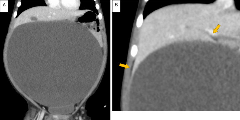
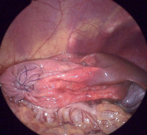
Case presentation: An 18-month-old girl presented with an enormous cyst occupying the entire abdomen. The beak sign on computed tomography revealed the hepatic origin of the cyst. The cyst was decompressed through the umbilicus, which was opened by the three-triangular-skin-flap technique, thus creating a working space that enabled laparoscopic surgery. The cyst was excised en bloc together with the attached hepatic parenchyma.
Conclusions: Giant simple hepatic cysts occupying the entire abdomen are rare in children. Of 14 reported cases, only 1 underwent laparoscopic treatment. We have herein reported another case of a giant simple hepatic cyst in which the beak sign on imaging and the three-triangular-skin-flap umbilical opening technique were useful for its diagnosis and laparoscopic excision, respectively. Complete excision is desirable because there is a possibility of recurrence or other diseases that require total removal, including hydatid cysts and mesenchymal hamartomas.
Keywords: Children; Enormous abdominal cyst; Laparoscopic excision; Simple hepatic cysts.
© 2022. The Author(s).
Conflict of interest statement
The authors declare that they have no competing interests.
Figures


Similar articles
-
Hepatic Hydatid Cyst Misdiagnosed as Simple Cyst: A Case Report.Am J Case Rep. 2020 Aug 2;21:e923281. doi: 10.12659/AJCR.923281. Am J Case Rep. 2020. PMID: 32740645 Free PMC article.
-
Traumatic rupture of a non-parasitic simple hepatic cyst presenting as an acute surgical abdomen: Case report.Int J Surg Case Rep. 2019;65:87-90. doi: 10.1016/j.ijscr.2019.10.051. Epub 2019 Oct 28. Int J Surg Case Rep. 2019. PMID: 31698200 Free PMC article.
-
Complete laparoscopic excision of a hepatic cyst and omentopexy in a Persian cat.JFMS Open Rep. 2018 Dec 16;4(2):2055116918817631. doi: 10.1177/2055116918817631. eCollection 2018 Jul-Dec. JFMS Open Rep. 2018. PMID: 30574339 Free PMC article.
-
Laparoscopic Excision of Congenital Hepatic Cysts in the Pediatric Population: A Case Series and Literature Review.J Laparoendosc Adv Surg Tech A. 2016 Jun;26(6):493-7. doi: 10.1089/lap.2016.0115. Epub 2016 May 5. J Laparoendosc Adv Surg Tech A. 2016. PMID: 27149195 Review.
-
Laparoscopic excision of the cystic lymphangioma occurred in the lesser omentum: report of a case and review of literature.Surg Laparosc Endosc Percutan Tech. 2009 Feb;19(1):e11-4. doi: 10.1097/SLE.0b013e31818a8a9b. Surg Laparosc Endosc Percutan Tech. 2009. PMID: 19238048 Review.
References
LinkOut - more resources
Full Text Sources
Research Materials
