. 2022 May 18;33(5):969-981.
doi: 10.1021/acs.bioconjchem.2c00167. Epub 2022 May 6.

One-Pot, One-Step Synthesis of Drug-Loaded Magnetic Multimicelle Aggregates

Affiliations

One-Pot, One-Step Synthesis of Drug-Loaded Magnetic Multimicelle Aggregates

Chang Soo Kim et al. Bioconjug Chem. .

Abstract

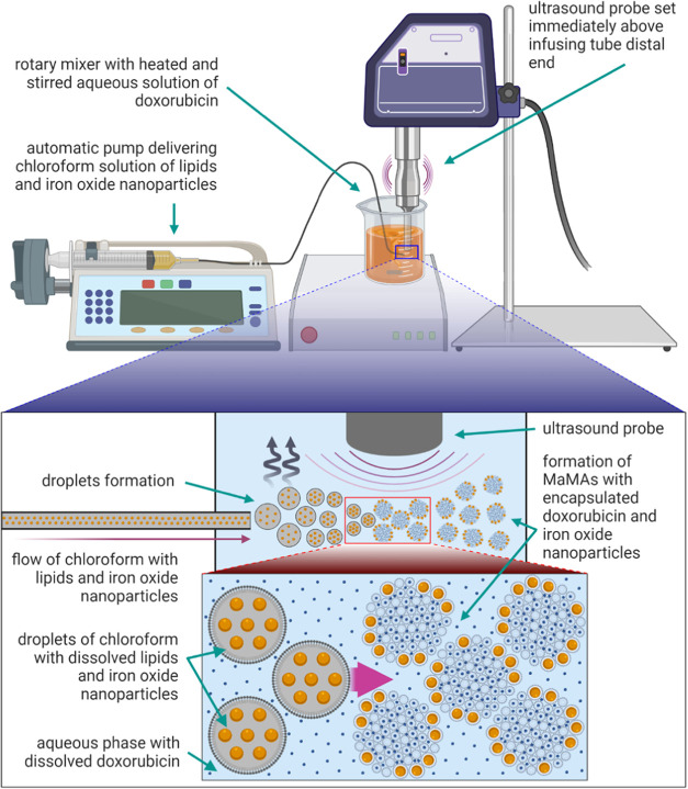
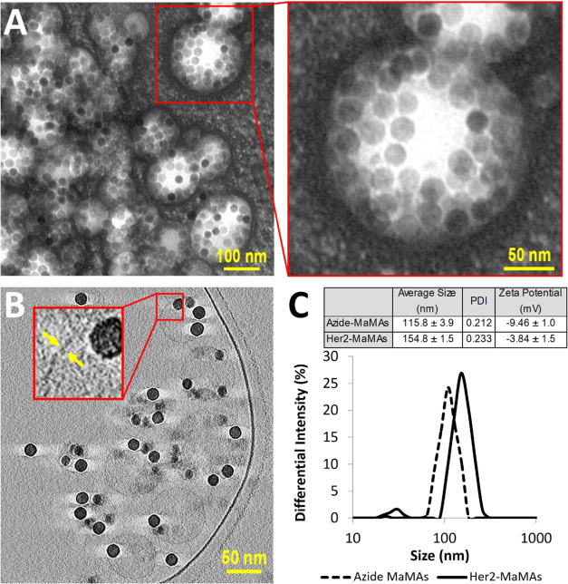
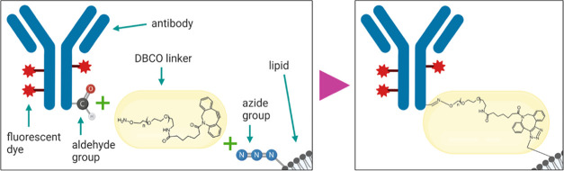
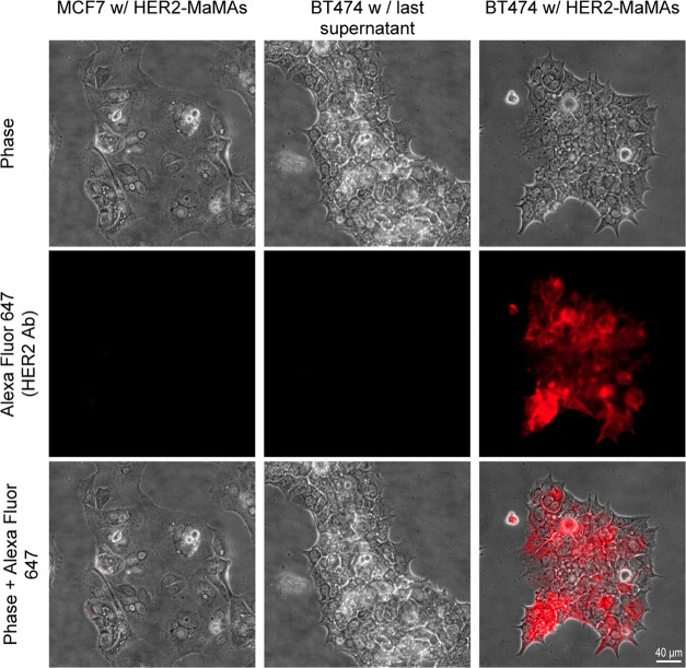
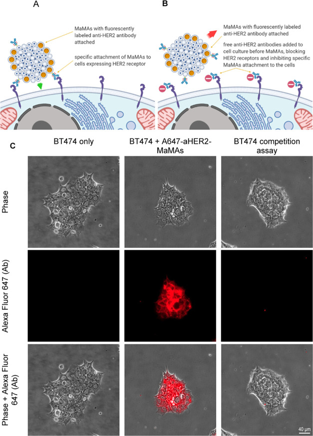
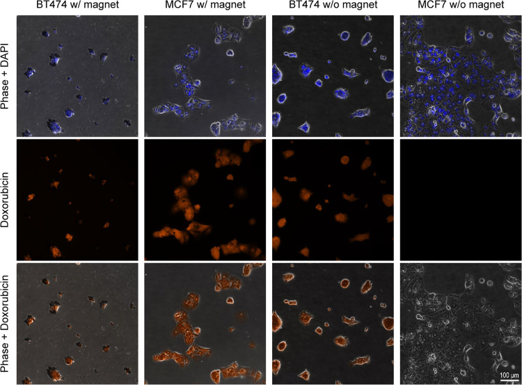
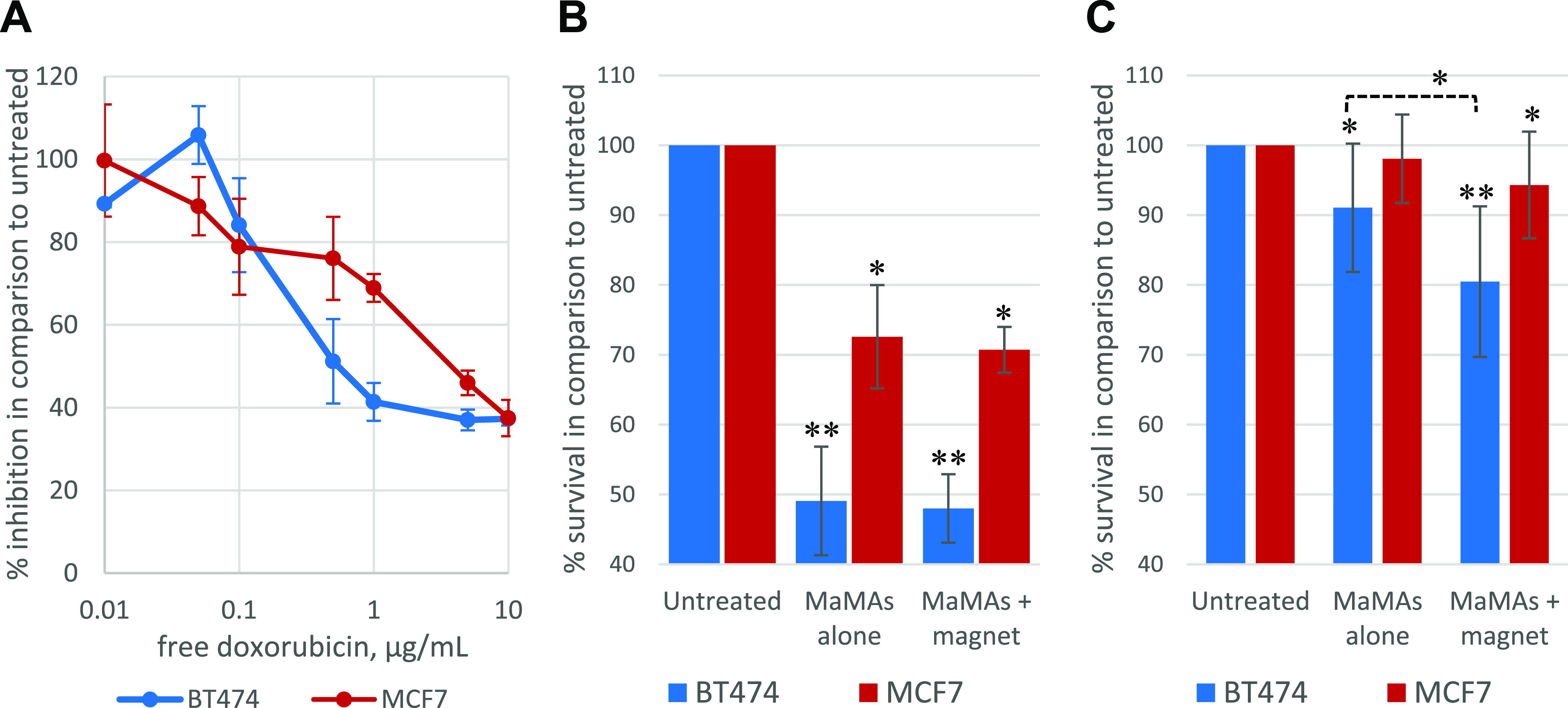
Lipid-based formulations provide a nanotechnology platform that is widely used in a variety of biomedical applications because it has several advantageous properties including biocompatibility, reduced toxicity, relative ease of surface modifications, and the possibility for efficient loading of drugs, biologics, and nanoparticles. A combination of lipid-based formulations with magnetic nanoparticles such as iron oxide was shown to be highly advantageous in a growing number of applications including magnet-mediated drug delivery and image-guided therapy. Currently, lipid-based formulations are prepared by multistep protocols. Simplification of the current multistep procedures can lead to a number of important technological advantages including significantly decreased processing time, higher reaction yield, better product reproducibility, and improved quality. Here, we introduce a one-pot, single-step synthesis of drug-loaded magnetic multimicelle aggregates (MaMAs), which is based on controlled flow infusion of an iron oxide nanoparticle/lipid mixture into an aqueous drug solution under ultrasonication. Furthermore, we prepared molecular-targeted MaMAs by directional antibody conjugation through an Fc moiety using Cu-free click chemistry. Fluorescence imaging and quantification confirmed that antibody-conjugated MaMAs showed high cell-specific targeting that was enhanced by magnetic delivery.


Conflict of interest statement

The authors declare the following competing financial interest(s): M.Z. is an employee of Imagion Biosystems. J.D.H. is a member of the Scientific Advisory Board, Imagion Biosystems.

Figures

Figure 1
Figure 1
Schematic of a one-pot, one-step synthesis of drug-loaded magnetic lipid-based formulations. The laboratory setup (top) and an outline of the formation process. Magnetic multimicelle aggregates (MaMAs) were synthesized by controlled flow infusion of an IONPs/lipid mixture into an aqueous solution of doxorubicin under ultrasonication.
Figure 2
Figure 2
(A) TEM images of MaMAs obtained using negative staining with 2% uranyl acetate. (B) Cross-sectional cryo-EM images of MaMAs. Individual IONPs (25 nm in diameter) can be clearly seen within spherical structures. Note the absence of a visible lipid bilayer, indicating that these structures are not liposomes. (C) Size (DLS, intensity) and ζ-potentials of MaMAs before and after conjugation with trastuzumab.
Figure 3
Figure 3
Schematic of fluorescently labeled trastuzumab antibody conjugation to azide-functionalized MaMAs through the bifunctional DBCO-PEG-aminooxy linker using Cu-free click chemistry. This approach utilizes mild oxidation of a carbohydrate moiety on the antibody’s Fc portion to form aldehyde groups.
Figure 4
Figure 4
Optical microscopy images of (from left to right) HER2 MCF7 cells after incubation with A647-aHER2-MaMAs; HER2+ BT474 incubated with the supernatant collected after the last washing step following synthesis of A647-aHER2-MaMAs (a control for free residual Alexa Fluor 647-labeled trastuzumab antibodies); and HER2+ BT474 cells after incubation with A647-aHER2-MaMAs. Note the lack of a fluorescence signal from HER2+ BT474 cells incubated with the supernatant; this indicates that the fluorescence after incubation with A647-aHER2-MaMAs is associated with the nanoparticles rather than with residual free antibodies. The images were acquired using a Zeiss Axio Observer.Z1m microscope equipped with a Hamamatsu ORCA-ER camera (Bridgewater, NJ) under a 40× objective lens. Fluorescence images were obtained with BP 640/30 nm excitation and BP 690/50 nm emission filters. Scale bar is 40 μm.
Figure 5
Figure 5
Blocking assay with HER2+ BT474 cells. Schematic of the study: (A) targeted MaMAs conjugated with fluorescently labeled trastuzumab antibodies (A647-aHER2-MaMAs) binding to HER2 positive cells; (B) blocking assay in which HER2 receptors are blocked by preincubation with free trastuzumab that precludes subsequent binding of A647-aHER2-MaMAs. (C) Optical microscopy images of (from left to right) BT474 cells alone (untreated control); BT474 A647-aHER2-MaMAs (BT474 cells labeled with A647-aHER2-MaMAs); and BT474 blocking assay where BT474 cells were preincubated with free trastuzumab antibodies before labeling with A467-aHER2-MaMAs. The images were acquired using a Zeiss Axio Observer.Z1m microscope equipped with a Hamamatsu ORCA-ER camera (Bridgewater, NJ) under a 40× objective lens. Fluorescence images were obtained with BP 640/30 nm excitation and BP 690/50 nm emission bandpass filters. Scale bar is 40 μm.
Figure 6
Figure 6
HER2+ BT474 and HER2 MCF7 cells after incubation with aHER2-DOX-MaMAs at 37 °C with or without a permanent magnet: (top row) combined phase and DAPI images; (middle row) fluorescence images of doxorubicin; and (bottom row) combined phase and doxorubicin images. The images were acquired using a Zeiss Axio Observer.Z1m microscope equipped with a Hamamatsu ORCA-ER camera (Bridgewater, NJ) under a 40× objective lens. Fluorescence images were obtained with BP 550/25 nm excitation and BP 605/70 nm emission filters for doxorubicin detection and G 365 nm excitation, BP 445/50 emission filters for DAPI. Scale bar is 100 μm for all images.
Figure 7
Figure 7
Antiproliferative effect in BT474 and MCF7 cells produced: (A) by free doxorubicin after continuous incubation with various drug concentrations for 72 h (n = 3); (B) after 72 h of continuous incubation with aHER2-DOX-MaMAs at 0.35 μg/mL free doxorubicin equivalent concentration (n = 3); (C) after 2.5 h of total incubation with aHER2-DOX-MaMAs at 0.0875 μg/mL free doxorubicin equivalent concentration (n = 11); in panel (C) a magnet was applied only during the first 30 min of incubation with the aHER2-DOX-MaMAs.

References

    1. Filipczak N.; Pan J.; Yalamarty S. S. K.; Torchilin V. P. Recent advancements in liposome technology. Adv. Drug Delivery Rev. 2020, 156, 4–22. 10.1016/j.addr.2020.06.022. - DOI - PubMed
    1. Ahmed K. S.; Hussein S. A.; Ali A. H.; Korma S. A.; Lipeng Q.; Jinghua C. Liposome: composition, characterisation, preparation, and recent innovation in clinical applications. J. Drug Target. 2019, 27, 742–761. 10.1080/1061186X.2018.1527337. - DOI - PubMed
    1. Jensen G. M.; Hodgson D. F. Opportunities and challenges in commercial pharmaceutical liposome applications. Adv. Drug Delivery Rev. 2020, 154–155, 2–12. 10.1016/j.addr.2020.07.016. - DOI - PubMed
    1. Al-Jamal W. T.; Kostarelos K. Liposomes: from a clinically established drug delivery system to a nanoparticle platform for theranostic nanomedicine. Acc. Chem. Res. 2011, 44, 1094–1104. 10.1021/ar200105p. - DOI - PubMed
    1. Carter K. A.; Shao S.; Hoopes M. I.; Luo D.; Ahsan B.; Grigoryants V. M.; Song W.; Huang H.; Zhang G.; Pandey R. K.; et al. Porphyrin-phospholipid liposomes permeabilized by near-infrared light. Nat. Commun. 2014, 5, 354610.1038/ncomms4546. - DOI - PMC - PubMed

Publication types