. 2022 May 6;17(5):e0267668.
doi: 10.1371/journal.pone.0267668. eCollection 2022.

Alternative methods for skeletal maturity estimation with the EOS scanner-Experience from 934 patients

Affiliations

Alternative methods for skeletal maturity estimation with the EOS scanner-Experience from 934 patients

Ádám Tibor Schlégl et al. PLoS One. .

Abstract

Background: Hand-wrist bone age assessment methods are not possible on typical EOS 2D/3D images without body position modifications that may affect spinal position. We aimed to identify and assess lesser known bone age assessment alternatives that may be applied retrospectively and without the need for extra imaging.

Materials and methods: After review of 2857 articles, nine bone age methods were selected and applied retrospectively in pilot study (thirteen individuals), followed by evaluation of EOS images of 934 4-24-year-olds. Difficulty of assessment and time taken were recorded, and reliability calculated.

Results: Five methods proved promising after pilot study. Risser 'plus' could be applied with no difficulty in 89.5% of scans (836/934) followed by the Oxford hip method (78.6%, 734/934), cervical (79.0%, 738/934), calcaneus (70.8%, 669/934) and the knee (68.2%, 667/934). Calcaneus and cervical methods proved to be fastest at 17.7s (95% confidence interval, 16.0s to 19.38s & 26.5s (95% CI, 22.16s to 30.75s), respectively, with Oxford hip the slowest at 82.0 s (95% CI, 76.12 to 87.88s). Difficulties included: regions lying outside of the image-assessment was difficult or impossible in upper cervical vertebrae (46/934 images 4.9%) and calcaneus methods (144/934 images, 15.4%); position: lower step length was associated with difficult lateral knee assessment & head/hand position with cervical evaluation; and resolution: in the higher stages of the hip, calcaneal and knee methods.

Conclusions: Hip, iliac crest and cervical regions can be assessed on the majority of EOS scans and may be useful for retrospective application. Calcaneus evaluation is a simple and rapidly applicable method that may be appropriate if consideration is given to include full imaging of the foot.


Conflict of interest statement

The authors have declared that no competing interests exist.

Figures
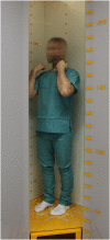
Fig 1
Fig 1. Patient position inside the EOS 2D/3D scanner.
Fig 2
Fig 2
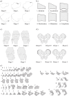
(a-e). Pictorial illustration of five bone age estimation methods. (a) Calcaneus, (b) Cervical, (c) Knee, (d) Risser Plus and (e) Oxford Hip methods. See S1 Fig for more in depth description of each method. (Reproduced from O’Sullivan et al. [17], creative commons license https://creativecommons.org/licenses/by/4.0/legalcode).
Fig 3
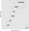
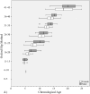
Fig 3. Distribution of chronological ages at each bone age stage with calcaneus method.
Gender is shown at each stage.
Fig 4
Fig 4. Distribution of chronological ages at each bone age stage with cervical method.
Gender is shown at each stage.
Fig 5
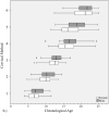
Fig 5. Distribution of chronological ages at each bone age stage with knee method.
Gender is shown at each stage.
Fig 6
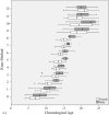
Fig 6. Distribution of chronological ages at each bone age stage with Oxford hip method.
Gender is shown at each stage.
Fig 7
Fig 7. Distribution of chronological ages at each bone age stage with Risser plus method.
Gender is shown at each stage.
Fig 8
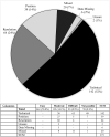
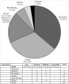
Fig 8. Difficulties reported during image assessment of the calcaneus method.
Difficulties were grouped as those due to technical difficulties e.g. landmark(s) lying outside of the edge of the image; resolution issues, insufficient resolution to assess image easily; positioning—landmark not visible, or not clearly visible due to limb rotation or shadowing; and if rater was ‘unsure’ due to problems with method description.
Fig 9
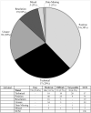
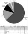
Fig 9. Difficulties reported during image assessment of the cervical method.
Difficulties were grouped as those due to technical difficulties e.g. landmark(s) lying outside of the edge of the image; resolution issues, insufficient resolution to assess image easily; positioning—landmark not visible, or not clearly visible due to limb rotation or shadowing; and if rater was ‘unsure’ due to problems with method description.
Fig 10
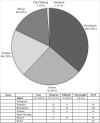
Fig 10. Difficulties reported during image assessment of the knee method.
Difficulties were grouped as those due to technical difficulties e.g. landmark(s) lying outside of the edge of the image; resolution issues, insufficient resolution to assess image easily; positioning—landmark not visible, or not clearly visible due to limb rotation or shadowing; and if rater was ‘unsure’ due to problems with method description.
Fig 11
Fig 11. Difficulties reported during image assessment of the Oxford hip method.
Difficulties were grouped as those due to technical difficulties e.g. landmark(s) lying outside of the edge of the image; resolution issues, insufficient resolution to assess image easily; positioning—landmark not visible, or not clearly visible due to limb rotation or shadowing; and if rater was ‘unsure’ due to problems with method description.
Fig 12
Fig 12. Difficulties reported during image assessment of the Risser plus method.
Difficulties were grouped as those due to technical difficulties e.g. landmark(s) lying outside of the edge of the image; resolution issues, insufficient resolution to assess image easily; positioning—landmark not visible, or not clearly visible due to limb rotation or shadowing; and if rater was ‘unsure’ due to problems with method description.
Fig 13
Fig 13. Timing data for each of the bone age methods.

References

    1. Dimeglio A, Canavese F. Progression or not progression? How to deal with adolescent idiopathic scoliosis during puberty. J Child Orthop. 2013;7:43–9. doi: 10.1007/s11832-012-0463-6 - DOI - PMC - PubMed
    1. Schlégl ÁT, Szuper K, Somoskeöy S, Than P. Three dimensional radiological imaging of normal lower-limb alignment in children. Int Orthop. 2015;39:2073–80. doi: 10.1007/s00264-015-2851-2 - DOI - PubMed
    1. Gheno R, Nectoux E, Herbaux B, Baldisserotto M, Glock L, Cotten A, et al.. Three-dimensional measurements of the lower extremity in children and adolescents using a low-dose biplanar X-ray device. Eur Radiol. 2012;22:765–71. doi: 10.1007/s00330-011-2308-y - DOI - PubMed
    1. Breen MA, Tsai A, Stamm A, Kleinman PK. Bone age assessment practices in infants and older children among Society for Pediatric Radiology members. Pediatr Radiol. 2016;46:1269–74. doi: 10.1007/s00247-016-3618-7 - DOI - PubMed
    1. Little DG, Song KM, Katz D, Herring JA. Relationship of peak height velocity to other maturity indicators in idiopathic scoliosis in girls. JBJS LWW. 2000;82:685–93. doi: 10.2106/00004623-200005000-00009 - DOI - PubMed

Publication types