. 2022 May 6;20(5):e3001635.
doi: 10.1371/journal.pbio.3001635. eCollection 2022 May.

Integrin-ECM interactions and membrane-associated Catalase cooperate to promote resilience of the Drosophila intestinal epithelium

Affiliations

Integrin-ECM interactions and membrane-associated Catalase cooperate to promote resilience of the Drosophila intestinal epithelium

Mohamed Mlih et al. PLoS Biol. .

Abstract

Balancing cellular demise and survival constitutes a key feature of resilience mechanisms that underlie the control of epithelial tissue damage. These resilience mechanisms often limit the burden of adaptive cellular stress responses to internal or external threats. We recently identified Diedel, a secreted protein/cytokine, as a potent antagonist of apoptosis-induced regulated cell death in the Drosophila intestinal midgut epithelium during aging. Here, we show that Diedel is a ligand for RGD-binding Integrins and is thus required for maintaining midgut epithelial cell attachment to the extracellular matrix (ECM)-derived basement membrane. Exploiting this function of Diedel, we uncovered a resilience mechanism of epithelial tissues, mediated by Integrin-ECM interactions, which shapes cell death spreading through the regulation of cell detachment and thus cell survival. Moreover, we found that resilient epithelial cells, enriched for Diedel-Integrin-ECM interactions, are characterized by membrane association of Catalase, thus preserving extracellular reactive oxygen species (ROS) balance to maintain epithelial integrity. Intracellular Catalase can relocalize to the extracellular membrane to limit cell death spreading and repair Integrin-ECM interactions induced by the amplification of extracellular ROS, which is a critical adaptive stress response. Membrane-associated Catalase, synergized with Integrin-ECM interactions, likely constitutes a resilience mechanism that helps balance cellular demise and survival within epithelial tissues.


Conflict of interest statement

The authors have declared that no competing interests exist.

Figures

Fig 1
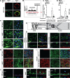
Fig 1. Diedel is required for enterocyte attachment to the basement membrane.
(A) Dissected Drosophila posterior midgut cross sections from control (WT, revertant [rev.]), Diedel mutant homozygote (dieΔ1/dieΔ1), and Diedel mutant trans-heterozygote (dieΔ1/dieΔ2) flies; stained with Phalloidin (Phall.; purple) and DAPI (blue). (B) Distance quantification between posterior midgut enterocyte nuclei (identified as large nuclei; DAPI; blue) and visceral muscle (Phalloidin) from above genotypes; WT, revertant [rev.] n = 80; dieΔ1/dieΔ1, n = 119; dieΔ1/dieΔ2, n = 58. (C) Phalloidin staining (Phall.; red) of midgut and hindgut visceral muscle dissected from WT, dieΔ1/dieΔ1, and dieΔ1/dieΔ2 flies. (D) Dissected posterior midgut cross sections from control flies (w1118; CGGal4) and flies with fat body attenuation of Diedel (w1118; CGGal4 / UAS-Die RNAi); stained with Phalloidin (Phall.; purple) and DAPI (blue). (E) Distance quantification between posterior midgut enterocyte nuclei (identified as large nuclei; DAPI; blue) and visceral muscle (Phalloidin) from above genotypes; w1118; CGGal4, n = 45; w1118; CGGal4 / UAS-Die RNAi, n = 85. (F) Die-V5 localization in dissected Drosophila midguts from controls (negative control; w1118) and w1118; DieP-Die-V5 / DieP-Die-V5 transgenic flies; stained with anti-V5 antibody (Die-V5; red) and DAPI (blue). (G) Schematic of Drosophila midgut organization (basement membrane). (H) Integrin Mys immunostaining of dissected posterior midguts from control (WT, revertant [rev.]) and Diedel mutant (dieΔ1/dieΔ1) flies; bottom panel represents cross-section (apical–basal polarity is highlighted with arrow); stained with anti-Mys (green), Phalloidin (Phall.; purple), and/or DAPI (blue).(I) Integrin Mys immunostaining of dissected posterior midguts from control flies (w1118; CGGal4) and flies with fat body attenuation of Diedel (w1118; CGGal4 / UAS-Die RNAi); top panels represent cross-sections (apical–basal polarity is highlighted with arrow); stained with anti-Mys (green), Phalloidin (Phall.; purple), and/or DAPI (blue). Data information: Data in panels B and E are presented as mean ± SD, n = 45–109. *P ≤ 0.05 (Student t test). The data underlying the graphs shown in Fig 1B and 1E can be found in S1 Data.
Fig 2
Fig 2. Diedel is a ligand for RGD-binding Integrins.
(A) Integrin Mys and anti-V5 immunostaining of dissected posterior midguts from w1118; DieP-Die-V5 / DieP-Die-V5; dieΔ1/dieΔ1 transgenic flies; bottom panel represents cross-section (apical–basal polarity is highlighted with arrow); stained with anti-Mys (green), anti-V5 antibody (Die-V5; red), and DAPI (blue). (B) Diedel protein sequence and topology with RGD domain highlight in red. (C) Die-V5 and DieRGE-V5 protein sequences and nucleotide sequence alignment; nucleotide and amino acid substitution highlighted in blue (top panel). Bottom panel; western blot with anti-V5 antibody of conditioned media (supernatant) from S2R+ cells either nontransfected (Mock), transfected with Die-V5, or transfected with DieRGE-V5. (D) Transfection of S2R+ cells (control; pmT empty) with Inflated Integrin (pMT Inflated), plated on conditioned media (supernatant) containing Mock (control), Die-V5, or DieRGE-V5; cells stained with Phalloidin (Phall.; white). (E) Quantification of “cell spreading” assay in S2R+ cells plated on various conditioned medias. The percentage of “spread” cells is shown, and bars indicate mean ± SD from n = 3 independent experiments (approximately 100 cells counted in each experiment). *P ≤ 0.05 (Student t test). (F) Integrin Mys immunostaining of dissected posterior midguts from control flies (w1118; CGGal4) and flies with fat body expression of either wild-type Diedel (w1118; CGGal4 / UAS-Die) or Diedel RGE (w1118; CGGal4 / UAS-DieRGE#1), stained with anti-Mys (green), Phalloidin (Phall.; red), and/or DAPI (blue). The data underlying the graphs shown in Fig 2E can be found in S1 Data.
Fig 3
Fig 3. Diedel prevents cell death spreading initiated by loss of Integrin–ECM interactions.
(A) Anti-V5 immunostaining of S2R+ cells before and after UV exposure (100 mJ/cm2); cells treated with Die-V5 conditioned media (supernatant); stained with anti-V5 antibody (Die-V5; red) and Phalloidin (Phall.; green). (B) Schematic representing coculture system to study cell death spreading in vitro. (B) UV-induced (100 mJ/cm2; +/− exposure) cell death spreading; quantified by percentage of Annexin V–positive cells. S2R+ cells treated with conditioned media (supernatant) containing Mock (control) or Die-V5. (C) Caspase (apoptosis)-induced (dsRNA targeting DIAP; dsRNA RFP as control) cell death spreading; quantified by percentage of Annexin V–positive cells. S2R+ cells treated with conditioned media (supernatant) containing Mock (control) or Die-V5. (D) Schematic of the ECM–Integrin–Talin complex; Cher (Cheerio) is an additional adaptor protein. (E) Talin immunostaining of dissected posterior midguts from control (WT, revertant [rev.]) and Diedel mutant (dieΔ1/dieΔ1 and dieΔ1/dieΔ2) flies; stained with anti-Talin (green) and DAPI (blue). (F) Talin immunostaining of dissected posterior midgut from control flies (w1118; CGGal4) and flies with fat body attenuation of Diedel (w1118; CGGal4 / UAS-Die RNAi); stained with anti-Talin (green), DAPI (blue), and Phalloidin (Phall.; red); bottom panel represents cross-section (apical–basal polarity is highlighted with arrow). (G) Integrin Mys and Caspase (cleaved Dcp-1) immunostaining of dissected posterior midguts from control (WT, revertant [rev.]) and Diedel mutant (dieΔ1/dieΔ1) flies; stained with anti-Mys (green), anti-Dcp-1 (Cleaved Dcp-1; red), and DAPI (blue). (H) Integrin Mys and Caspase (cleaved Dcp-1) immunostaining of dissected posterior midguts from control flies (w1118; CGGal4) and flies with fat body attenuation of Diedel (w1118; CGGal4 / UAS-Die RNAi); stained with anti-Mys (green), anti-Dcp-1 (Cleaved Dcp-1; red), and DAPI (blue). (I) Posterior midgut cross sections (apical–basal polarity is highlighted with arrow) from control (WT, revertant [rev.]) and Diedel mutant (dieΔ1/dieΔ1 and dieΔ1/dieΔ2) flies; stained with anti-cleaved Cas3 (green), DAPI (blue), and Phalloidin (Phall.; red). Strong staining of Cas3 (revealing cells detaching from the basement membrane) are highlighted by solid white arrowhead). Data information: Data in panels C and D are presented as mean ± SD, n = 3–5. *P ≤ 0.05 (Student t test). The data underlying the graphs shown in Fig 3C and 3D can be found in S1 Data.
Fig 4
Fig 4. Catalase associates with cellular membranes/ECM after stress.
(A) Mass spectrometry of gel-based pulldown analysis. Protein identification (gel elutions provided) after Die-V5 pulldown from S2R+ cells treated with control (Mock) or with Die-V5 conditioned media (supernatant); after stress (UV exposure; 100 mJ/cm2). Table represents Mass-Spec protein identification with percent protein coverage. (B) Mass spectrometry of gel-free pulldown analysis (Diedel-interacting proteins). Venn diagrams showing overlap of proteins after Die-V5 pulldown from S2R+ cells treated with control (Mock) or with Die-V5 conditioned media (supernatant); after stress (UV exposure; 100 mJ/cm2). The threshold for proteins included in the analysis was at least 2 different fragments identified/sequenced. (C) Histogram plotting percentage of protein enrichment in S2R+ cells treated with Die-V5 conditioned media after UV exposure. The analysis included only cytoplasmic and membrane proteins with at least 2 different fragments identified/sequenced. (D) Catalase and anti-V5 immunostaining of S2R+ cells after UV exposure (100 mJ/cm2); cells treated with Die-V5 conditioned media (supernatant) or Mock (control); stained with anti-V5 antibody (Die-V5; red) and anti-Catalase (green). Cells were not permeabilized (nonpermeabilized) with detergent in order to visualize membrane/ECM proteins. (E) Catalase localization in primary cells expressing endogenous catalase tagged with GFP (green) after UV exposure (100 mJ/cm2); cells treated with Die-V5 conditioned media (supernatant) or Mock (control); stained with anti-V5 antibody (Die-V5; red). (F) UV-induced (100 mJ/cm2; +/− exposure) cell death quantification by percentage of TUNEL-positive cells. S2R+ cells were treated with dsRNA control (RFP) or dsRNA Catalase (cat), then exposed to UV (100 mJ/cm2) and treated with Die-V5 conditioned media (supernatant) or Mock (control). Data information: Data in panel F are presented as mean ± SD, n = 8–14. *P ≤ 0.05 (Student t test). The data underlying the graphs shown in Fig 4B can be found in S1 Table, and the data underlying the graphs shown in Fig 4C and 4F can be found in S1 Data.
Fig 5
Fig 5. Catalase relocalization to membranes is integrated with cell death spreading and extracellular ROS after epithelial tissue damage.
(A) Catalase immunostaining of dissected posterior midguts from control (w1118, Ctrl.) flies with DSS treatment/feeding (4% DSS) or mock treatment; stained with anti-Catalase (green) and DAPI (blue). Flies were fed (or Mock treated) DSS for 2 days. Immunostaining was performed in both permeabilized (to visualize intracellular Catalase) and nonpermeabilized (to visualize extracellular Catalase) conditions. (B) Cat-GFP immunostaining in dissected midguts from control (Ctrl.) MARCM clones and “Undead” MARCM clones 5 days after heat shock. Clones are marked with nuclear RFP (red), endogenously GFP-tagged Catalase (Catalase-GFP/+) are marked with GFP (green). White arrows highlight membrane localized Catalase in adjacent (“neighbor”) cells of “undead” MARCM clones. (C) Catalase localization and membrane/ECM cysteine oxidation of dissected posterior midguts from w1118, Catalase-GFP / Catalase-GFP (endogenously tagged) flies; DSS treatment/feeding (4% DSS), or mock treatment, or cotreatment/feeding with a Catalase inhibitor (DSS + 3-AT); stained with anti-Catalase (green), anti-Cysteine (oxidized, Cys. Ox; red), and DAPI (blue). Immunostaining was performed in nonpermeabilized conditions to visualize membrane/ECM cysteine oxidation. Panels represent cross-sections (apical–basal polarity is highlighted with arrow). Flies were fed (or Mock treated) DSS or DSS + 3-AT for 2 days. ECM, extracellular matrix; MARCM, mosaic analysis with a repressible cell marker; ROS, reactive oxygen species.
Fig 6
Fig 6. Membrane-associated Catalase is necessary to promote ECM preservation / repair and prevent cell death spreading in concert with Diedel–Integrin–ECM interactions.
(A) Membrane/ECM cysteine oxidation of dissected posterior midguts from w1118 (Ctrl.) flies; DSS treatment/feeding (4% DSS), or mock treatment, or cotreatment/feeding with a Catalase inhibitor (DSS + 3-AT), as well as 48 hours posttreatment (recovery after DSS feeding); stained with anti-Cysteine (oxidized, Cys. Ox; red) and DAPI (blue). Immunostaining was performed in nonpermeabilized conditions to visualize membrane/ECM cysteine oxidation. Flies were fed (or Mock treated) DSS or DSS + 3-AT for 2 days. (B) Integrin Mys and Caspase (cleaved Dcp-1) immunostaining of dissected posterior midguts from control (w1118) flies after DSS treatment/feeding (4% DSS), or mock treatment, or cotreatment/feeding with a Catalase inhibitor (DSS + 3-AT); stained with anti-Mys (green), anti-Dcp-1 (Cleaved Dcp-1; red), and DAPI (blue). Flies were fed (or Mock treated) DSS or DSS + 3-AT for 2 days. (C) Coculture system to study cell death spreading in vitro. UV-induced (100 mJ/cm2) cell death spreading; quantified by percentage of TUNEL-positive cells. S2R+ cells treated with conditioned media (supernatant) containing Mock (control) or Die-V5, and Catalase (3-AT), (D) Images of TUNEL immunostaining from cocultured S2R+ cells; UV-induced (100 mJ/cm2) cell death spreading. Cells treated with conditioned media (supernatant) containing Mock (control) or Die-V5, and Catalase inhibitor (3-AT). TUNEL (nuclear, green), DAPI (blue), phalloidin (membrane, red). Data information: Data in panel C are presented as mean ± SD, n = 9. *P ≤ 0.05 (Student t test). The data underlying the graphs shown in Fig 6C can be found in S1 Data.

Update of

Similar articles

Cited by

References

    1. Smirnova L, Harris G, Leist M, Hartung T. Cellular resilience. ALTEX. 2015;32:247–60. doi: 10.14573/altex.1509271 - DOI - PubMed
    1. Pazarentzos E, Bivona TG. Adaptive stress signaling in targeted cancer therapy resistance. Oncogene 2015 3445. 2015;34(45):5599–5606. doi: 10.1038/onc.2015.26 - DOI - PMC - PubMed
    1. Jamieson AM. Host resilience to emerging coronaviruses. Future Virol. 2016;11(7):529–534. doi: 10.2217/fvl-2016-0060 - DOI - PMC - PubMed
    1. Lee KZ, Lestradet M, Socha C, Schirmeier S, Schmitz A, Spenlé C, et al.. Enterocyte Purge and Rapid Recovery Is a Resilience Reaction of the Gut Epithelium to Pore-Forming Toxin Attack. Cell Host Microbe. 2016;20:716–30. doi: 10.1016/j.chom.2016.10.010 - DOI - PubMed
    1. Li X, Rommelaere S, Kondo S, Lemaitre B. Renal Purge of Hemolymphatic Lipids Prevents the Accumulation of ROS-Induced Inflammatory Oxidized Lipids and Protects Drosophila from Tissue Damage. Immunity. 2020;52:374–387.e6. doi: 10.1016/j.immuni.2020.01.008 - DOI - PubMed

Publication types

LinkOut - more resources