Modelling of magnetic microbubbles to evaluate contrast enhanced magnetomotive ultrasound in lymph nodes - a pre-clinical study
- PMID: 35522781
- PMCID: PMC10996324
- DOI: 10.1259/bjr.20211128
Modelling of magnetic microbubbles to evaluate contrast enhanced magnetomotive ultrasound in lymph nodes - a pre-clinical study
Abstract
Objectives: Despite advances in MRI the detection and characterisation of lymph nodes in rectal cancer remains complex, especially when assessing the response to neoadjuvant treatment. An alternative approach is functional imaging, previously shown to aid characterisation of cancer tissues. We report proof of concept of the novel technique Contrast-Enhanced Magneto-Motive Ultrasound (CE-MMUS) to recover information relating to local perfusion and lymphatic drainage, and interrogate tissue mechanical properties through magnetically induced deformations.
Methods: The feasibility of the proposed application was explored using a combination of experimental animal and phantom ultrasound imaging, along with finite element analysis. First, contrast-enhanced ultrasound imaging on one wild type mouse recorded lymphatic drainage of magnetic microbubbles after bolus injection. Second, tissue phantoms were imaged using MMUS to illustrate the force- and elasticity dependence of the magnetomotion. Third, the magnetomechanical interactions of a magnetic microbubble with an elastic solid were simulated using finite element software.
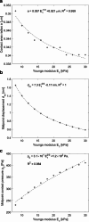
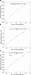
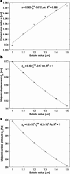
Results: Accumulation of magnetic microbubbles in the inguinal lymph node was verified using contrast enhanced ultrasound, with peak enhancement occurring 3.7 s post-injection. The magnetic microbubble gave rise to displacements depending on force, elasticity, and bubble radius, indicating an inverse relation between displacement and the latter two.
Conclusion: Combining magnetic microbubbles with MMUS could harness the advantages of both techniques, to provide perfusion information, robust lymph node delineation and characterisation based on mechanical properties.
Advances in knowledge: (a) Lymphatic drainage of magnetic microbubbles visualised using contrast-enhanced ultrasound imaging and (b) magnetomechanical interactions between such bubbles and surrounding tissue could both contribute to (c) robust detection and characterisation of lymph nodes.
Figures


Similar articles
-
Contrast enhanced magneto-motive ultrasound in lymph nodes - modelling and pre-clinical imaging using magnetic microbubbles.Annu Int Conf IEEE Eng Med Biol Soc. 2022 Jul;2022:194-197. doi: 10.1109/EMBC48229.2022.9871876. Annu Int Conf IEEE Eng Med Biol Soc. 2022. PMID: 36086230
-
High Frame Rate Contrast-Enhanced Ultrasound Imaging for Slow Lymphatic Flow: Influence of Ultrasound Pressure and Flow Rate on Bubble Disruption and Image Persistence.Ultrasound Med Biol. 2019 Sep;45(9):2456-2470. doi: 10.1016/j.ultrasmedbio.2019.05.016. Epub 2019 Jul 3. Ultrasound Med Biol. 2019. PMID: 31279503
-
Dynamic visualization of lymphatic channels and sentinel lymph nodes using intradermal microbubbles and contrast-enhanced ultrasound in a swine model and patients with breast cancer.J Ultrasound Med. 2010 Dec;29(12):1699-704. doi: 10.7863/jum.2010.29.12.1699. J Ultrasound Med. 2010. PMID: 21098840
-
Prospects of perfusion contrast-enhanced ultrasound (CE-US) in diagnosing axillary lymph node metastases in breast cancer: a comparison with lymphatic CE-US.J Med Ultrason (2001). 2024 Oct;51(4):587-597. doi: 10.1007/s10396-024-01444-w. Epub 2024 Apr 20. J Med Ultrason (2001). 2024. PMID: 38642268 Free PMC article. Review.
-
Ultrasound Contrast: Gas Microbubbles in the Vasculature.Invest Radiol. 2021 Jan;56(1):50-61. doi: 10.1097/RLI.0000000000000733. Invest Radiol. 2021. PMID: 33181574 Review.
Cited by
-
The use of ultrasound in colonic and perianal diseases.Curr Opin Gastroenterol. 2023 Jan 1;39(1):50-56. doi: 10.1097/MOG.0000000000000891. Epub 2022 Nov 3. Curr Opin Gastroenterol. 2023. PMID: 36504036 Free PMC article. Review.
References
-
- Barth A, Craig PH, Silverstein MJ . Predictors of axillary lymph node metastases in patients with T1 breast carcinoma . Cancer 1997. ; 79: 1918 – 22 . - PubMed

