

Imaging of bone marrow pitfalls with emphasis on MRI
- PMID: 35522786
- PMCID: PMC9975530
- DOI: 10.1259/bjr.20220063
Imaging of bone marrow pitfalls with emphasis on MRI
Abstract
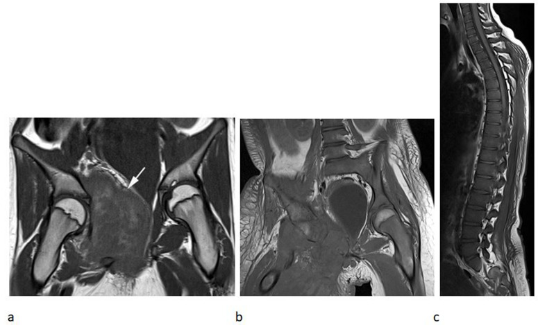
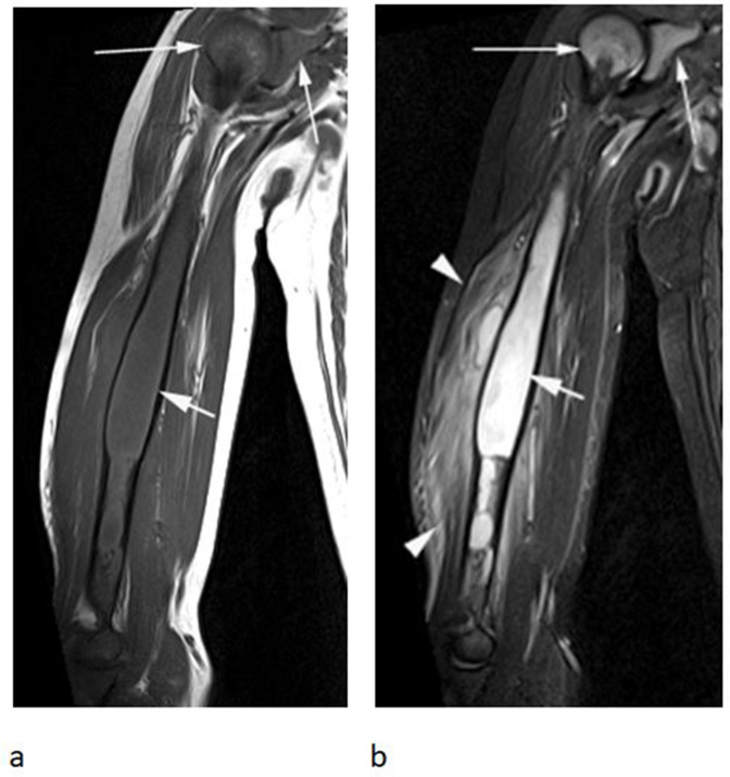
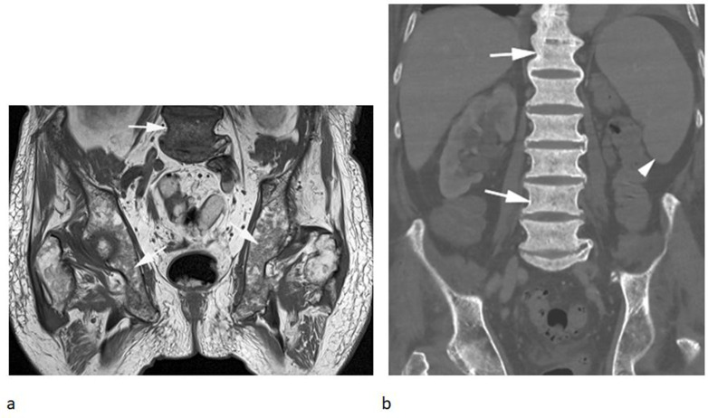
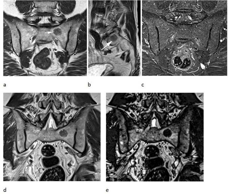
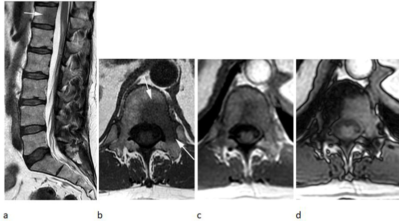
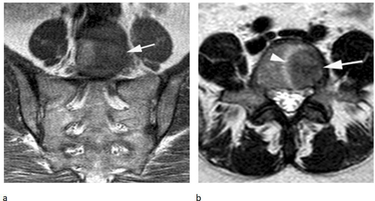
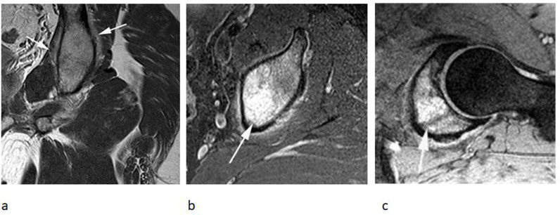
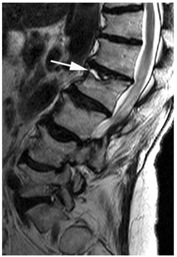
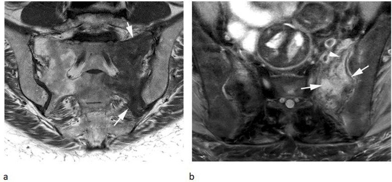
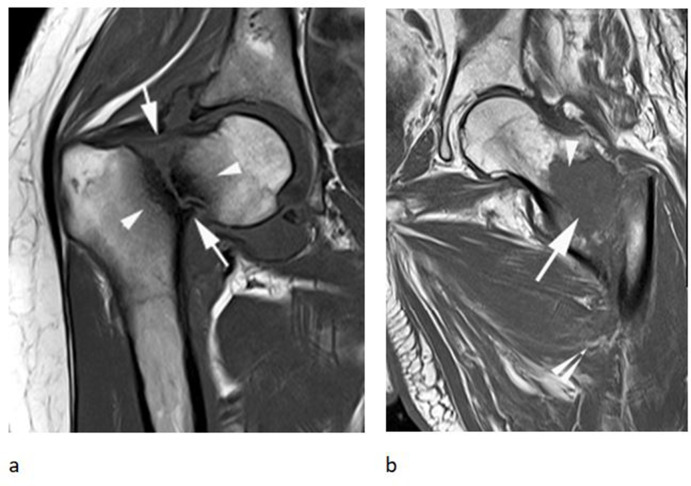
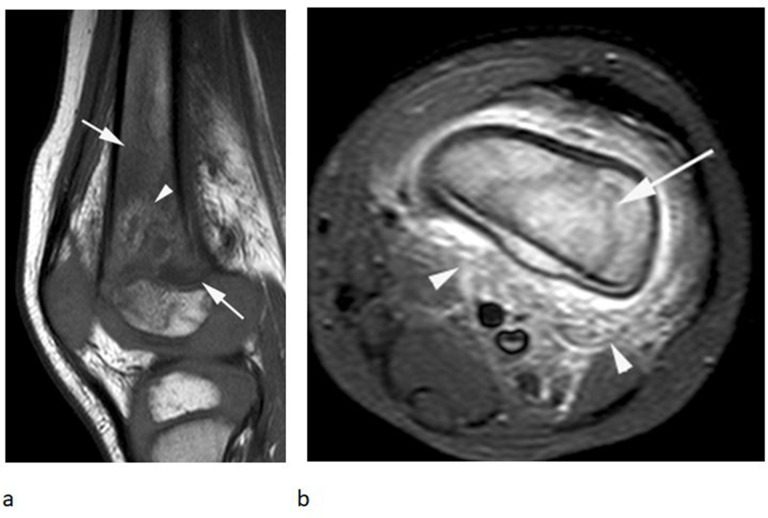
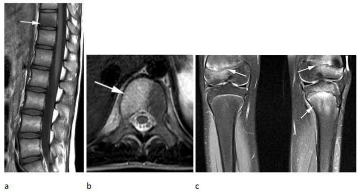
Normal marrow contains both hematopoietic/red and fatty/yellow marrow with a predictable pattern of conversion and skeletal distribution on MRI. Many variations in normal bone marrow signal and appearances are apparent and the reporting radiologist must differentiate these from other non-neoplastic, benign or neoplastic processes. The advent of chemical shift imaging has helped in characterising and differentiating more focal heterogeneous areas of red marrow from marrow infiltration. This review aims to cover the MRI appearances of normal marrow, its evolution with age, marrow reconversion, variations of normal marrow signal, causes of oedema-like marrow signal, and some common non-neoplastic entities, which may mimic marrow neoplasms.
Figures


References
-
- Bracken J, Nandurkar D, Radhakrishnan K, Ditchfield M. Normal paediatric bone marrow: magnetic resonance imaging appearances from birth to 5 years: MRI appearance of normal marrow from 0-5 years. J Med Imaging Radiat Oncol 2013; 57: 283–91. - PubMed
Publication types
MeSH terms
LinkOut - more resources
Full Text Sources
Medical
